Showing posts sorted by date for query aliya. Sort by relevance Show all posts

Saturday, July 27, 2024

EXERCISE IN GLAUCOMA: TO DO OR NOT TO DO?


DR. ALIYA

P.G. SCHOLAR

DEPT OF ILAJ-BIT-TADBEER

STATE UNANI MEDICAL COLLEGE AND HOSPITAL

PRAYAGRAJ, INDIA


Controversy exists among the patients and the healthcare industry about whether exercise, especially certain exercises such as yoga, is useful or harmful for glaucoma patients.

Many articles have shown the positive and negative sides of exercises on glaucoma status.

PROS OF EXERCISE:

Studies have shown that aerobic exercises are particularly useful in glaucoma patients. These exercises improve circulation in the brain and therefore, are useful for the eye, especially in glaucoma patients who have vascular anomalies contributing to glaucomatous damage.

A prominent glaucoma expert, Dr. Robert Ritch recommends 45 minutes of aerobic exercise 3-4 times per week. Walking, swimming, biking, or working out on stationary machines lowers intraocular pressure (IOP) and improves blood circulation to the eye and brain.

A study by Lee et al reported that increased walking, greater time spent doing moderate-to-vigorous physical activity, and more time spent in non-sedentary activity were associated with slower rates of VF loss in a treated population of patients with glaucoma, with an additional 5000 daily steps or 2.6 hours of non-sedentary physical activity decreasing the average rate of VF loss by approximately 10%. [1]

A study by Janicijevic et al reported that low-intensity aerobic exercise had a lowering effect on IOP, being the beneficial effect more accentuated and prolonged in the High-fit group (IOP reduction compared to baseline lasted 30 minutes) than in the Low-fit group (IOP was only reduced at 6 minutes of exercise compared to baseline). [2] 

Ramulu and associates have also found that substantial reductions in physical activity and walking were noted with greater levels of VF loss. [3] 

In experiments on mice, Chrysostomou and collegues reported that exercise almost completely reversed age-related vulnerability of the optic nerve to injury such that exercised aged mice had a similar functional response to injury as non-exercised young (3-month-old) mice. Exercise also abrogated injury-induced astrocytic gliosis and macrophage activation in the aged retina. These data suggest that the known benefits of exercise also extend to the visual system and support further investigation of physical activity as a means of protecting against injury, dysfunction, and degeneration in the aging eye. [4] 

A meta-analysis has found that mild-intensity aerobic exercises are beneficial because of their diverse mechanisms in glaucoma patients. They can help in the transient reduction of IOP and have a beneficial effect on glaucoma severity and progression. [5] 

CONS OF EXERCISE:

A large study from the UK Biobank Eye and Vision Consortium did not find a significant association between exercise and glaucoma. Higher overall physical activity (PA) level and greater time spent in moderate and vigorous exercise were not associated with glaucoma status but were associated with thicker mGCIPL. Associations with IOP were modest and inconsistent. Despite the well-documented acute reduction in IOP after PA, there was no evidence that high levels of habitual PA are associated with glaucoma status or IOP in the general population. [6] 

Certain yoga postures are especially harmful for glaucoma patients. Studies have shown downward facing dog (adho mukha svanasana), standing forward bend (uttanasana), Plow (halasana), and legs up the wall (viparita karani) are especially harmful in glaucoma patients. A study found that the above mentioned four poses raised IOP in both the control group and the glaucoma patients, with the greatest increases associated with downward-facing dog. Once the subjects returned to a seated position, they were tested immediately and 10 minutes later. The IOP returned to baseline levels at these time points.

A study by Liu et al found that arterioles show increased stiffness with aging and it cannot be reversed with exercise.[7] 

CONCLUSION:

It can be inferred from the above studies that light aerobic exercises are useful, but heavy exercises like weight-lifting and certain yoga postures are deleterious for glaucoma patients and should be avoided.


REFERENCES:

  1. Moon Jeong Lee, Jiangxia Wang, David S. Friedman, Michael V. Boland, Carlos G. De Moraes, Pradeep Y. Ramulu, Greater Physical Activity Is Associated with Slower Visual Field Loss in Glaucoma. Ophthalmology, Volume 126, Issue 7,2019,Pages 958-964.
  2. Janicijevic, D., Redondo, B., Jiménez, R., Garcia-Ramos, A., & Vera, J. (2022). The intraocular pressure lowering-effect of low-intensity aerobic exercise is greater in fitter individuals: a cluster analysis. Research in Sports Medicine, 32(1), 86–97.
  3. Pradeep Y. Ramulu, Eugenio Maul, Chad Hochberg, Emilie S. Chan, Luigi Ferrucci, David S. Friedman, Real-World Assessment of Physical Activity in Glaucoma Using an Accelerometer, Ophthalmology, Volume 119, Issue 6, 2012, Pages 1159-1166, https://doi.org/10.1016/j.ophtha.2012.01.013.
  4. Chrysostomou V, Kezic JM, Trounce IA, Crowston JG. Forced exercise protects the aged optic nerve against intraocular pressure injury. Neurobiol Aging. 2014 Jul;35(7):1722-5. doi: 10.1016/j.neurobiolaging.2014.01.019. Epub 2014 Jan 23. PMID: 24524967.
  5. Gildea, David MB BCh BAO; Doyle, Aoife MB, BCh; O’Connor, Jeremy MB, BCh. The Effect of Exercise on Intraocular Pressure and Glaucoma. Journal of Glaucoma 33(6):p 381-386, June 2024. | DOI: 10.1097/IJG.0000000000002411.
  6. Madjedi KM, Stuart KV, Chua SYL, Ramulu PY, Warwick A, Luben RN, Sun Z, Chia MA, Aschard H, Wiggs JL, Kang JH, Pasquale LR, Foster PJ, Khawaja AP; Modifiable Risk Factors for Glaucoma Collaboration and the UK Biobank Eye and Vision Consortium. The Association of Physical Activity with Glaucoma and Related Traits in the UK Biobank. Ophthalmology. 2023 Oct;130(10):1024-1036. doi: 10.1016/j.ophtha.2023.06.009. Epub 2023 Jun 17. PMID: 37331483; PMCID: PMC10913205.
  7. Liu C, Kobayashi T, Shiba T, Hayashi N (2022) Effects of aging and exercise habits on blood flow profile of the ocular circulation. PLoS ONE 17(4): e0266684.


Thursday, March 19, 2020

PRIMARY OPEN ANGLE GLAUCOMA


INTRODUCTION:


Glaucoma is characterized by the common characteristic feature of retinal ganglion cell loss and optic nerve degeneration. There are different methods to classify glaucomas. It can be categorized on the basis of etiology (Primary vs. secondary), duration (Acute vs. chronic), and anatomy of the anterior chamber angle (open vs. closed).


Primary Open Angle Glaucoma (POAG) is defined as a chronic, slowly progressive, optic neuropathy with characteristic patterns of optic nerve damage and visual field loss. It is a subset of the glaucomas characterized by an open, normal appearing anterior chamber angle and raised intraocular pressure (IOP), with no other underlying disease.

World-wide this type of glaucoma is the most prevalent. The global prevalence of glaucoma among individuals in the 40-80 years age group is around 3.54%. POAG accounts for 3.05% and primary angle closure glaucoma (PACG) 0.5% of the total prevalence. According to the WHO, nearly 5.2 million people are blind world-wide due to glaucoma. Of these, congenital glaucoma accounts for 200,000; POAG 3 million and 2 million from PACG.

There are many theories to explain the development of POAG. These include the mechanical, vascular, biochemical, genetic and intraluminal pressure theories. However, it is not known which etiology might be active in any particular patient. It is often assumed that multiple mechanisms could be responsible for glaucomatous damage in an individual. 

CLINICAL FEATURES:

POAG is usually insidious in onset, slowly progressive, and painless. It is often a bilateral, asymmetric disorder. The condition is called the “silent thief of sight” as central visual acuity is relatively unaffected until late in the disease. A number of risk factors for the development of POAG are known. These include intra-ocular pressure, increasing age, race, decreased central corneal thickness (CCT) and positive family history.

Intra-ocular pressure:

Based on population studies the normal mean IOP is 15.5 mmHg with a standard deviation of 2.6 mmHg. This makes the range of mean normal IOP between 10-21 mmHg. However, it is known that IOP in the general population is skewed to the right. A number of abnormalities in IOP measurement have been presented over the last few years. The role of normal diurnal fluctuation of IOP has shown that IOP measurements can be biased depending upon the time of measurement. This is more obvious in glaucoma patients where IOP may fluctuate by more than 8-10 mmHg (compared to 5 mmHg in normal eyes). 


Central corneal thickness:

CCT was found to be a powerful predictor for the development of POAG in OHTS. The relative risk of POAG increased 81% for every 40 μ thinning of cornea. Both OHTS and the European Glaucoma Prevention Study found that the risk of developing POAG was greater in eyes with CCT <555 μ compared with eyes having CCT of 588 μ or greater. Another study has reported that patients with thinner corneas tend to have more severe glaucoma even on initial examination and have a higher risk of progression. The actual IOP can be overestimated on Goldman applanation tonometry in eyes with thicker corneas, whereas an underestimation may happen in eyes with less than average CCT. Refractive surgery can alter the corneal biomechanics and corneal thickness, thus resulting in falsely low IOP readings. However, in the presence of corneal edema IOP tends to be underestimated and is overestimated when measured over corneal scars due to the increased rigidity of fibrous tissue.

Thicker corneas resist indentation during tonometry leading to erroneously high IOP readings. Conversely, in thin corneas the measured IOP can be falsely low. This can affect the diagnosis of POAG. Corneal thickness may be measured (pachymetry) by optical and ultrasonic methods.

Average corneal thickness, determined by optical and ultrasonic pachymetry, is approximately 530-545 µ in eyes without glaucoma. Central corneal thickness (CCT) is increased in patients with ocular hypertension (OHT). 

Other confounding factors in IOP measurement are corneal steepness (Steeper corneas resist indentation) and a mechanical factor called corneal hysteresis. Certain new instruments such as dynamic contour tonometry and Ocular Response Analyzer are able to overcome some of these factors.


Optic disc assessment and visual field loss:

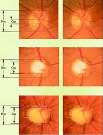
Abnormal cupping or an increase in the cup‑to‑disc ratio (CDR) is frequently associated with glaucoma suspects. Stereoscopic evaluation of the optic discs has shown a Gaussian distribution of the mean CDR at 0.4 with only an approximately 5% normal population having CDRs of 0.7 or more. A difference of 0.2 between the two eyes should be viewed with suspicion. Such a finding is present in only 1% of the normal population.

It is still not very clear whether structural changes (obvious on optic disc assessment and other tests) develop earlier than functional changes (seen on perimetric tests). However, careful periodic evaluation of the optic disc and visual field is vital in the follow-up of glaucoma patients. Stereophotographs of the optic discs are ideal to preserve the records of the patient for future reference. However, computerized digital images or even hand drawings may suffice, with the relevant details marked in the drawing. Stereophotographic documentation or computerized imaging of the disc are useful as they enhance the clinician's ability to detect subtle changes over time. 

A number of mechanical and vascular signs are found in glaucoma patients. These include:

Mechanical signs
Vascular signs
Large optic cup
Disc hemorrhage
Asymmetrical cups
Nasal displacement of vessels
Progressive enlargement of cup
Baring of circumlinear vessels
Narrowing/notching of rim
Tortuosity of retinal vessels on
the disc
Vertical elongation of cup

RNFL loss

Exposed lamina cribrosa (laminar dot sign)

Peripapillary atrophy

RNFL= Retinal nerve fiber layer

Particular attention should be paid to the neuroretinal rim (NRR). The rim is broadest inferiorly among all quadrants, followed by the superior, nasal, and temporal rims (ISNT‑rule). The loss of inferior or superior NRR leads to a vertical elongation of the cup and loss of the ISNT‑rule leading to a suspicion of glaucoma. It needs to be highlighted that ISNT rule is only applicable to normal sized discs.


Visual field loss should correlate with the appearance of the optic disc. Significant discrepancies in the pattern of field loss and optic nerve damage warrant additional investigation. As perimetry remains a highly subjective test, on an average, 3 VF assessments should be done in the 1st year to detect an overall change in mean deviation of 4 dB over 2 years in a patient with average VF variability. Progressive VF loss is the hallmark sign which separates a true pathology from a glaucoma suspect.

Gonioscopy:

Assessment of anterior chamber angles is imperative for the diagnosis of POAG. The clinician should be well versed with the procedure of gonioscopy. Angles which are more than Grade 2 are regarded as open. If peripheral anterior synechiae are present, the extent of PAS should be recorded. 


Increasing age:

In a number of landmark studies increasing age was noted as a significant risk factor. In the Baltimore Eye Study the risk of glaucoma increased significantly in patients above 80 years of age. In the Collaborative Initial Glaucoma Treatment Study (CIGTS) visual field defects were more common in patients above 60 years of age compared to those below 40 years.

Family history:

The lifetime risk for first-degree relatives of affected individuals to develop open angle glaucoma is 22% when compared with a 2% risk in controls. it is likely both monogenic and polygenic. Instances where a single gene causes glaucoma (monogenic) have emerged (e.g., the myocilin and optineurin genes), but in other instances glaucoma is much more likely the result of multiple genes (polygenic).

Unique susceptibility genes and genetic variants also likely play a significant role in glaucoma.

TREATMENT:

Management of open angle glaucoma has to be tailored for every patient. Initial treatment usually consists of pharmacological therapy. Prostaglandin analogues and beta blockers are the preferred agents to initiate treatment. Depending upon various factors drugs can be changed or added. Compliance is an essential factor to be considered when target IOP is not achieved. Some clinicians prefer to perform laser procedures (Argon Laser Trabeculoplasty) or even implantation of glaucoma drainage devices (GDDs) as initial procedures. However, usually GDDs are used in advanced cases. Trabeculectomy or Minimally Invasive Glaucoma Surgery (MIGS) can also be utilized in certain patients. Glaucoma filtering surgery is usually performed in the situation of uncontrolled IOP despite maximally tolerable medical therapy. MIGS is usually combined with cataract surgery. In case there is poor visual potential and ocular discomfort (painful, blind eye) the eye may be treated with cyclodestructive procedures. A number of laser and surgical methods are available. These include diode laser cyclophotocoagulation, micropulse-trans-scleral cyclophotocoagulation and endocyclophotocoagulation. Surgical techniques such as cyclodialysis are not commonly in vogue now.
 
Progressive glaucomatous optic atrophy

Thursday, July 25, 2019

CENTRAL CORNEAL THICKNESS AND GLAUCOMA


Guest author

ALIYA

Ajmal Khan Tibbiya College
Aligarh
India 


INTRODUCTION

  • According to the 11th century Arab scholar, Abu Al-Hassan Tabarri, a firm or hard eye was an indication of glaucoma. Over the years intraocular pressure (IOP) has become the bedrock in the positive and negative association with glaucoma. On one hand IOP is regarded as a surrogate factor in the assessment of glaucoma, while on the other, certain conditions such as ocular hypertension and normal-tension glaucoma question the importance of this marker to diagnose glaucoma.
  • There are a number of instruments developed over the years to measure IOP. However, despite all our advances, Goldmann Applanation Tonometry (GAT) continues to be labeled as the “gold standard” to assess this parameter. Unfortunately, GAT is fraught with a number of errors such as improper technique; patient factors such as cooperation during measurement and tight neckties; size of the semi-circles (Wider menisci cause falsely higher pressure estimates. Improper vertical alignment will also lead to a falsely high IOP estimate.); corneal factors such as corneal thickness, curvature and astigmatism; calibration errors and error due to prolonged contact.
  • GAT is dependent on a standard central corneal thickness (CCT), leading to errors in IOP measurement if the CCT is beyond the standard values (explained later). Unfortunately, the mean CCT in healthy human eyes varies by ethnoracial characteristics. This produces controversial confounding factors in the measurement of IOP by GAT.
  • Apart from being a factor in IOP measurement, CCT is also regarded as a risk factor for development of glaucoma and also associated with glaucoma severity and progression.
  • Most clinical practice guidelines do recommend evaluation of CCT in the initial assessment of all glaucoma patients. This post takes a look at CCT and how it affects glaucoma diagnosis and management.

CCT AND OCULAR HYPERTENSION:


  • The Ocular Hypertension Treatment Study (OHTS) has shown that thin CCT is the most important predictive factor for the development of primary open angle glaucoma (POAG).
  • However, there are indications that the significance of CCT as a risk factor for glaucoma could have been overestimated in OHTS.
  • OHTS assessed patients who had high IOP at baseline (without other glaucoma characteristics), this led to a selection bias towards individuals who had thicker CCT.
  • 25% patients had CCT above 600µ, so a significant number of participants in OHTS had corrected IOP of less than 21 mmHg.
  • Conversely, individuals with thin CCT would have their IOP overestimated during eligibility screening.
  • These factors would lead to overestimation of the significance of CCT as a risk factor for the development of glaucoma.
  • Although these limitations are important, CCT has been established as an important factor to consider in the risk stratification of OHT patients.
  • The OHTS prediction model was independently validated in the European Glaucoma Prevention Study (EGPS) and also in the pooled OHTS-EPGS data.
  • Every 40µ decrease in CCT was associated with a two-fold increase in the risk of developing glaucoma over 5 years.
  • CCT measurement would minimize the overtreatment of OHT patients with thick corneas.
  • In patients with thin CCT and borderline IOP, initiating treatment can be considered keeping in view other relevant risk factors.
  • It needs to be emphasized that IOP alone (even when adjusted for CCT) is not a reliable screening tool for POAG.

CCT AND GLAUCOMA


  • The role of CCT in the clinical management of patients with established glaucoma is less well recognized (compared to OHT).
  • Prevalence of POAG was significantly higher in individuals with thin (compared to normal or thick) CCTs in the Los Angeles Latino Eye Study.
  • Studies in West Africa, East Asia and China did not show any significant difference in CCT in glaucomatous versus non-glaucomatous eyes.
  • Studies have shown eyes with NTG are associated with thinner CCT than eyes with POAG and normal eyes.
  • Pseudoexfoliative glaucoma and PACG have been found to be associated with thin corneas in some studies but refuted in others.
  • It is controversial whether any significant association exists between CCT and glaucoma progression or glaucoma severity.
  • Early Mainfest Glaucoma Trial (EMGT) did show thinner CCT to be associated with glaucoma progression.
  • However, other landmark glaucoma trials such as Advanced Glaucoma Intervention Study (AGIS), Collaborative Initial Glaucoma Treatment Study (CIGTS) and Collaborative Normal Tension Glaucoma Study (CNTGS) did not analyze the impact of CCT on glaucoma.
  • A few other recent studies have not found any significant association between CCT and glaucoma.

POSSIBLE MECHANISMS FOR THE ASSOCIATION BETWEEN CCT AND GLAUCOMA


(1) Tonometry artifact:

  • GAT is based on an average CCT of 500µ.
  • Ehlers cannulated the anterior chamber to obtain true IOP levels and found the most accurate GAT results were obtained with CCT of 520µ.
  • Every 100µ of deviation in CCT would produce an error of 7 mmHg in IOP measurement.
  • A thick cornea causes an artefactually increase IOP reading, while a thin cornea produces an artefactually reduced IOP reading.
  • Corneal compensated IOP is either independent or weakly dependent on CCT.
  • Certain tonometers such as Ocular Response Analyzer (ORA) and Corvis tonometer are able to compensate for CCT.
  • Other tonometers such as Dynamic Contour Tonometry and Transpalpebral Tonometry have their own drawbacks.
  • Linear correction factors for IOP based on CCT have been proposed, however based on mathematical models, the relationship between CCT and IOP is non-linear and complex.
  • So far no adequately validated correction algorithm for GAT measurements has been provided.

(2) Biological risk factor:

  • While certain studies have shown increased movement of lamina cribrosa in response to IOP changes in individuals with thin CCTs, others have refuted this.
  • Another hypothesis suggests that a thin cornea increases the exposure of the trabecular meshwork to oxidative damage (a study reported an inverse correlation between CCT and partial pressure of oxygen in the anterior chamber).

(3) Genetic associations:

  • CCT is often inherited, with heritability estimates for CCT ranging from 88-95%.
  • Apparently a number of genes are responsible for inheritance of CCT.
  • Again, while there are studies suggesting the role of mutations and single nucleotide polymorphisms, others did not find any genetic correlation between CCT and POAG.

GLAUCOMA TREATMENT


  • Ocular hypotensive medications have been shown to alter corneal thickness.
  • Most studies have reported an association between the use of topical prostaglandin analogues (PGA), including latanoprost and tafluprost, and a significant decrease in CCT, possibly by modifying the extracellular matrix.
  • However, there are other studies which did not find any correlations between PGA use and CCT.
  • Topical beta-blockers have also been reported to cause reversible increase in CCT.
  • Topical carbonic anhydrase inhibitors can cause irreversible corneal decompensation and increased corneal thickness in eyes with underlying corneal disease. However, this effect is not seen in healthy corneas.
  • Conversely, there appears to be an effect of corneal thickness on the response of glaucoma treatment.
  • The OHTS reported that thicker CCT was associated with significantly less decrease in IOP in response to anti-glaucoma treatment.
  • In a study of Selective Laser Trabeculoplasty for POAG and OHT, thinner corneas were associated with comparatively higher IOP reduction.

VARIABILITY IN CCT

  • CCT measurements vary with race, sex, age and other environmental factors, which may confound the relationship between glaucoma and corneal thickness.
  • African (518-533µ), Indian (511-514µ), Mongolian (495-514µ) and Japanese (517-532µ) individuals were found to have thinner corneas compared to white (542-558µ), Hispanic (547µ), Korean (554µ) and Chinese (540-542µ) persons in population-based studies.
  • Racial differences in CCT would need to be considered in the interpretation of IOP and may explain the higher rates of glaucoma in certain populations.
  • Wang et al. found that the variation in CCT accounted for upto 29.4% of the increased risk of glaucoma seen among African-American persons in a large multiethnic population in Northern California.
  • In the OHTS, African-American ethnicity was predictive of POAG in univariate analysis, though it was not significant in the multivariate analysis after inclusion of CCT in the model, suggesting the increased susceptibility of this group of individuals to glaucoma can be partially explained by their thin CCT.
  • Most epidemiological studies found men to have thicker corneas.
  • CCT varies with age, with a small but significant inverse relationship between CCT and age (range: 2-10microns per decade). This is comparable to longitudinal data from OHTS (6microns per decade).
  • It can be surmised that it would take 20 years for the change in CCT to be clinically significant and to warrant remeasuring.
  • CCT is also influenced by environmental factors. Dry eyes and long term contact lens wear are associated with a decrease in CCT, whereas occupation indoors and sleep are associated with an increase in CCT.
  • The use of various devices which use different modalities to measure CCT has also resulted in variable results of CCT.
  • Most landmark glaucoma studies such as OHTS, EMGTS and EGPS used ultrasound pachymetry.
  • There are other instruments also being used to measure CCT. These include: Scheimpflug imaging, optical coherence tomography, specular microscopy and scanning slit topography.
  • It is ideal to use the same device for the same patient for every CCT measurement.

CORNEAL BIOMECHANICS

  • Corneal biomechanics may have a more significant impact on IOP measurement errors than CCT which is merely a corneal dimension and not a biomechanical property.
  • CCT is regarded as a correlate of corneal rigidity, but this is only applicable to structurally normal Cornea.
  • An edematous Cornea has lower corneal rigidity and therefore, despite increased thickness will have lower GAT readings.
  • Conversely, a scarred cornea, although thinner, has increased rigidity and will demonstrate artefactually higher IOP readings.
  • Eyes undergoing corneal cross linking show higher IOP on an average 12 months after the procedure, apparently due to higher corneal rigidity.
  • Measurements of Corneal biomechanical properties, such as corneal hysteresis, corneal resistance factor and corneal constant factor can be obtained by the Ocular Response Analyzer and the Corvis ST tonometer.
  • These parameters may be better correlated with overall globe biomechanics than CCT and may help to explain the susceptibility of some optic nerve heads to damage as a result of IOP variations.


NERVE GROWTH FACTORS FOR TREATMENT OF GLAUCOMA

  Neurotrophin-based therapies, such as recombinant human nerve growth factor (rhNGF), are promising candidates for non-IOP based treatments...