Thursday, July 6, 2023

ASTROCYTES


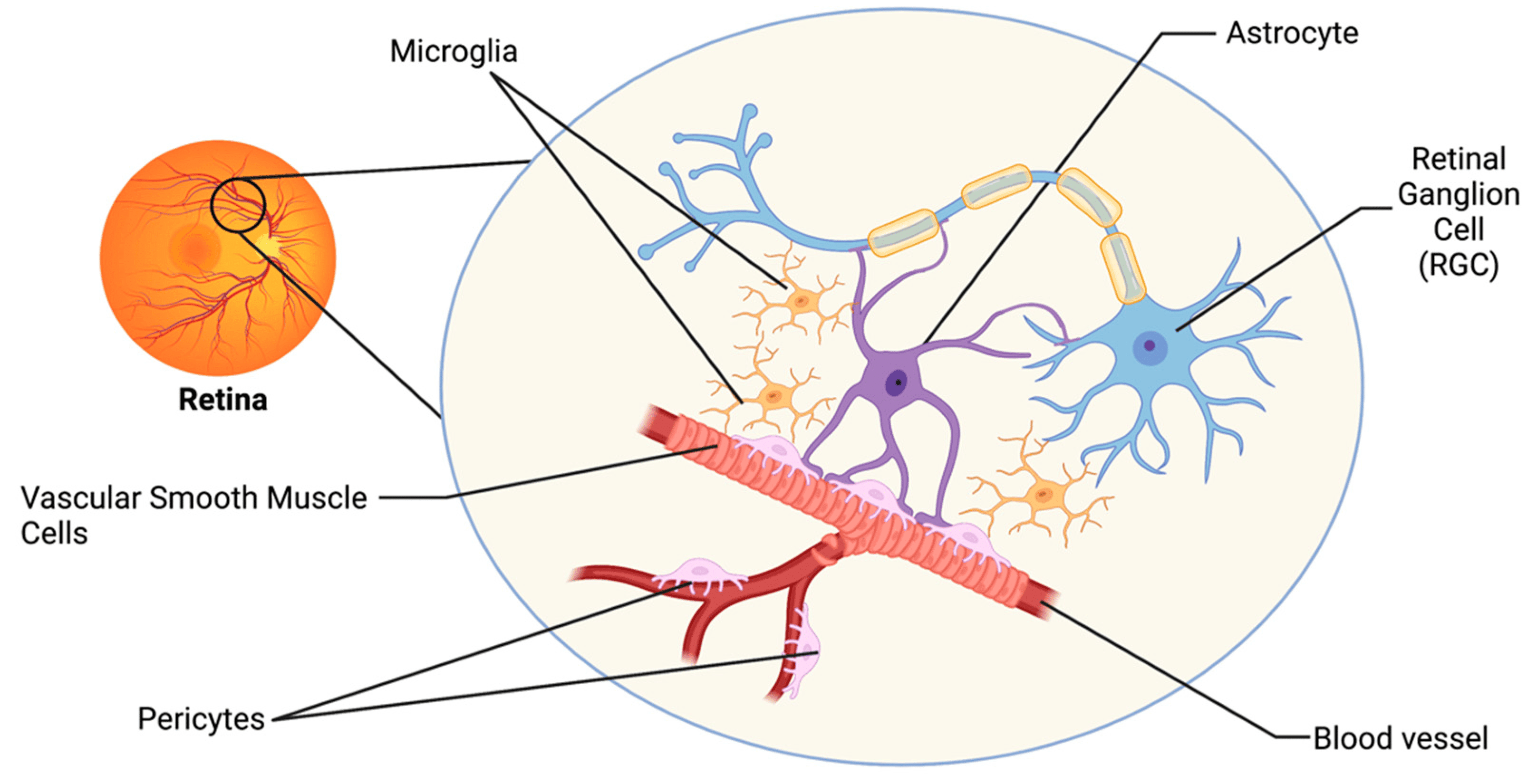
Astrocytes are the major glial cell type in the non-myelinated optic nerve head (ONH) in most mammals and provide cellular support functions to the axons while interfacing between connective tissue surfaces and surrounding blood vessels.

Astrocytes form a mesh-like network on the surface of the retina and tightly contact blood vessels and retinal ganglion cell (RGC) axons.


ONH astrocytes are responsible for the normal maintenance of the extra-cellular matrix (ECM) in normal eyes. These cells are sensitive to mechanical or ischemic factors and are important for the maintenance of retinal ganglion physiology.

1A astrocytes provide structural support for the axons, with type 1B cells providing a physiological interface between the vitreous connective and vascular tissues.

In the normal ONH, astrocytes are considered to be quiescent.


In the lamina cribrosa, astrocytes form lamellae, oriented perpendicular to the axons surrounding a core of fibroelastic extracellular matrix.


Astrocytes supply energy substrate to axons in the optic nerve and maintain extracellular pH and ion homeostasis in the periaxonal space. Sodium channels in astrocytes participate in Na+ homeostasis, providing a path for Na+ entry into the cytoplasm.

In astrocytes, voltage-gated calcium channels deliver Ca2+ into the cytoplasm and participate in generation of glial Ca2+ signals.

Astrocytes maintain the scant periaxonal ECM consisting of glycoproteins, such as laminin and proteoglycans.

Astrocytes express a wide variety of growth factors and receptors, many of which serve as trophic and survival factors for neurons.

Astrocytes are the cells responsible for many pathological changes in the glaucomatous optic nerve head (ONH).

Astrocytes become reactive in response to injury or disease and participate in the formation of a glial scar, which does not support axonal survival or growth.

The major hallmarks of a reactive astrocyte are an enlarged cell body and a thick network of processes with increased expression of GFAP and vimentin.

Reactive astrocytes increase expression of various cell surface molecules that play important roles in cell–cell recognition and in cell adhesion to substrates, as well as various growth factors, cytokines, and receptors. Reactive astrocytes express many new ECM proteins such as laminin, tenascin C, and proteoglycans.

Reactive astrocytes in the glaucomatous ONH are large rounded cells with many thick processes which expresses increased amounts of GFAP, vimentin, and HSP27.

Recent evidence suggests that optic nerve head astrocytes, which have long been recognized as important components of the optic nerve head, may underlie this process and be central to the initiation of glaucomatous optic atrophy. These cells probably have a direct toxic effect on the RGC axons.

In glaucoma, reactive astrocytes have been shown to migrate from the cribriform plates into the nerve bundles and synthesize neurotoxic mediators such as nitric oxide (NO) and TNF-α, which may be released near axons causing neuronal damage.

Reactive astrocytes in the ONH express large amounts of elastin, leading to elastotic degeneration of the ECM in glaucoma and loss of resiliency and deformability in response to elevated IOP.

ONH astrocytes may offer neuroprotection in the optic nerve by releasing glutathione (GSH) and antioxidant enzymes to eliminate the products of chronic oxidative stress that may be contributing to the progression of neurodegeneration in POAG.

Astrocyte dysfunction could disrupt axoplasmic transport and initiate the changes in cribrosal physiology that are said to be secondary to the mechanical effects of raised IOP or to ischemic damage secondary to optic disc hypoperfusion.

This hypothesis implies that significant disturbances of astrocyte metabolism may predispose to axon loss and initiate changes in cribrosal structure. Thus, the collapse of cribrosal beams, rather than initiating axon loss, may be as much the result of astrocyte fallout.


Saturday, July 1, 2023

BIOMECHANICS and GLAUCOMA


Biomechanics is a newly developed interdisciplinary subject which applies mechanical principles and technology to biological systems.


The normal human sclera is composed from the outside to the inside of episclera, scleral stroma and lamina fascia. The episclera contains an irregular arrangement of interwoven small collagen fibers. The scleral stroma is composed of dense collagen fiber bundles containing many collagenous fibrils which lie parallel to each other on the outer surface, and which interlace and fuse together on the inner surface. The lamina fascia is composed of smaller collagen fiber bundles. Thus, collagen plays a major role in maintaining the structure, function and the biomechanical properties of the sclera.

The biomechanical theory of glaucoma proposes that optic nerve head (ONH) biomechanics may explain how IOP-induced stress and strain (a measure of tissue deformation) of the load bearing tissues of the ONH (sclera and lamina cribrosa/LC) influence their physiology and pathophysiology, and of the adjunctive tissues (astrocytes, glia, endothelial cells, vascular pericytes and their basement membranes) and the retinal ganglion cell (RGC) axons.

The biomechanical model of the disease proposes that IOP-induced mechanical strain on the ONH leads to a cascade of events eventually culminating in RGC dysfunction and apoptosis.


The ‘compliance’ (the inverse of stiffness) of the normal ONH in response to an acute elevation of IOP have been studied using techniques of increasing complexity. These include: X-ray photography of cadaveric non-human primate eyes with fine platinum wires inserted into the peripapillary sclera and optic disc, laser doppler velocimetry of normal human autopsy eyes, conventional histology of human eyes, 2D- and 3D-histo-morphometric reconstructions of post mortem normal monkey eyes. These reports were all consistent (to a greater or lesser degree) in finding a posterior movement of the optic disc surface in response to an acute elevation of IOP.

Finite element modeling is a computational tool for predicting how a complex biological tissue will behave under varying levels of load. Finite element modeling in monkey and human cadaver eyes have been used to study the mechanical response of the sclera to different levels of IOP.

The biomechanical model suggests that a given level of IOP-related stress may be physiologic or pathophysiologic depending on the individual ONH experiencing the stress.

Physiologic levels of IOP-related stress are assumed to be capable of inducing a broad spectrum of acute and chronic changes in all three ONH tissue types.

Pathophysiologic levels of IOP-related stress are assumed to induce pathologic changes in cell synthesis and tissue microarchitecture.

Scleral material properties, geometry, and thickness have significant effects on the biomechanical environment of the ONH.

There may be significant inter-individual differences in biomechanical behavior stemming from differences in the anatomy of the sclera.

The biomechanical environment within the ONH may play a role in RGC loss in glaucomatous optic neuropathy.

Biomechanically, it is hypothesized that IOP-induced mechanical strain on glial cells supporting ganglion cell axons eventually leads to apoptosis of the RGCs and the consequent loss of vision.

The “safe” level of IOP is patient specific, a difference that is likely due, at least in part, to differences in ONH geometry and biomechanical properties.

Strain represents the amount of stretching that a tissue undergoes, and stress is the force within the tissue per unit of tissue area. Strain is important since most research in mechanobiology suggests that cells respond to strain (deformation) rather than directly to stress. Stress is important because it determines the tendency of extracellular materials to fail (tear) as occurs at the periphery of the glaucomatous LC.


DEFERIPRONE IN GLAUCOMA MANAGEMENT

  Currently, lowering of intra-ocular pressure (IOP) remains the main therapeutic option for the treatment of glaucoma. However, studies hav...