Friday, May 20, 2022

PARASYMPATHETIC OR ANTI-CHOLINERGIC DRUGS


Parasympathetic or Cholinergic drugs mimic the action of acetylcholine (ACh), a neuro-transmitter at the postganglionic parasympathetic junction, as well as at other autonomic, somatic and central synapses.

Ach is synthesized by “Choline Acetyltransferase” enzyme and produces its effects by binding to cholinergic receptors at the effector site.


Cholinergic drugs act either through direct stimulation of cholinergic receptors or indirectly through inhibition of the “Cholinesterase”, thereby protecting endogenous Ach.

Cholinergics affect intra-ocular pressure (IOP) by an agonist-induced, muscarinic receptor-mediated contraction of the ciliary muscle.

There are two ways in which ciliary muscle contraction can affect aqueous outflow.

One, ciliary muscle contraction obliterates the intermuscular spaces within the ciliary muscle. This obstructs uveoscleral outflow.

Secondly, there is intimate relationship of the anterior tendons of the ciliary muscle bundles with the scleral spur, peripheral cornea, trabecular meshwork and inner wall of Schlemm’s canal. Ciliary muscle contraction results in an unfolding of the trabecular meshwork and widening of Schlemm’s canal, facilitating aqueous outflow from the anterior chamber through the meshwork into the canal lumen and thence into the venous collector channels and finally, the general venous circulation.

However, the facilitation of outflow via the trabecular meshwork (conventional pathway) more than compensates for the obstruction of the uveoscleral (unconventional) route. The net effect of ciliary muscle contraction, therefore, is to decrease IOP.

Another effect of ciliary muscle contraction is accommodative myopia by the cholinomimetic agents. This is a major drawback, especially in young individuals.

Induced miosis can be a problem for elderly patients with immature cataracts.

Muscarinic agents are of 2 types:

Directly acting= They stimulate the iris sphincter to cause miosis, and stimulate the ciliary muscle to increase outflow facility, decrease uveoscleral outflow and produce accommodative changes.

Examples of directly acting cholinergic agents include pilocarpine aceclidine, arecoline, acetyl-B-methylcholine.

Indirectly acting= They block cholinesterase (AChE) preventing metabolic inactivation of Ach released from parasympathetic nerve endings. When AChE is blocked, the local concentration of endogenously released Ach and its time of action are increased. This increases and prolongs the response of endogenous cholinergics.

Examples of indirectly acting agents include physostigmine, demecarium, echothiaphate, and isoflurophate.


PILOCARPINE

History and source:

It is an alkaloid produced from the leaflets of South American shrubs of the genus Pilocarpus (P. microphyllus). Pilocarpine was foirst introduced as an anti-glaucoma agent in 1877.

Pharmacology:

Pilocarpine acts by direct stimulation of muscarinic cholinergic receptors. It duplicates the muscarinic effects of Ach, but not its nicotinic effects.

Clinical pharmacology:

It produces miosis through contraction of the iris sphincter muscle, which pulls the iris root away from the trabecular meshwork in angle-closure glaucoma. It also causes ciliary muscle contraction, resulting in accommodation and increased tension on and opening of the trabecular meshwork spaces, facilitating aqueous humor outflow and lowering IOP in open-angle glaucoma.

Pharmacokinetics:

Pilocarpine penetrates the cornea well. Animal studies have shown that the cornea absorbs pilocarpine rapidly and then releases it slowly to the aqueous humor. Onset of miosis with a 1% solution is within 10-30 minutes. Maximum reduction in IOP occurs in 75 minutes when a solution is used. The duration of action for miosis is about 4-8 hours. The reduction in IOP lasts for 4-14 hours.

In brown eyed patients 4% solution maybe required for maximum effect, whereas in extremely dark individuals 8-10% solution is the most effective. The differences are due to the binding of the drug by pigment within the eye, making it unavailable to the relevant muscarinic receptors.

The drug is inactivated by tissues of the anterior segment of the eye, partly by reversible binding of the drug to tissues and also by enzymatic hydrolysis to the primary metabolite, pilocarpic acid.

Resistance to the IOP lowering effect may occur after prolonged use.

Therapeutic use:

Pilocarpine is generally used in a concentration of 0.5-4.0% aqueous solution four times per day. 

In acute angle closure it can be instilled 2-3 times over a 30-minute period.

Side effects and toxicity:

Transient stinging and burning are common. Conjunctival vascular congestion and true allergy are unusual. Prolonged use of pilocarpine may alter the conjunctival tissues to make subsequent glaucoma filtration surgery more likely to fail. Intraocular vascular congestion may occur in and aggravate uveitic conditions. Ciliary spasm, temporal or supraorbital headache and induced myopia may occur. Reduced visual acuity in dim illumination may occur in elderly with central lens opacities. Accelerated development of lens opacities is also reported. Retinal detachment in susceptible individuals may occur.

Sweating and gastro-intestinal over activity usually occurs in children. Over dosage can produce sweating, salivation, nausea, tremors, slowing of pulse and decreased blood pressure.

High-risk groups:

Pilocarpine is not advised in conditions where pupillary constriction and intraocular vascular congestion are undesirable, such as in acute iritis.

It should also be avoided in individuals prone for retinal detachment, severe asthma, or bronchial obstruction or acute infectious conjunctivitis or keratitis.

Caution should also be exercised when prescribing to children.


CARBACHOL

History and source:

Carbachol is a carbamyl ester of choline and synthesized in 1930s.

Pharmacology:

It has direct sympathomimetic actions as well as an indirect mechanism of action by inhibition of AChE.


Pharmaceutics:

Isopto Carbachol (0.75, 1.5, 2.25 and 3.0%) solution is administered every 8 hourly.

Miostat (0.01%) solution is used as intracameral injection during surgery.

Pharmacokinetics:

Carbachol is not lipid soluble and therefore, penetrates intact cornea poorly.

Maximum reduction of IOP occurs within 4 hours and lasts about 8 hours. Miosis lasts 4-8 hours.

Intracameral miostat produces maximal miosis within 5 minutes and lasts about 24 hours.

Therapeutic use:

0.75-3.0% solution is used 3 times daily.

Side effects and toxicity:

Same as Pilocarpine.

High risk groups:

It is contra-indicated in the presence of iritis. Caution is advised in the presence of corneal abrasion to prevent excessive intraocular penetration.

Caution is also advised in the presence of acute cardiac failure, bronchial asthma, active peptic ulcer, hyperthyroidism, gastrointestinal spasm, urinary tract obstruction, Parkinson’s disease, recent myocardial infarction, systemic hypertension or hypotension.
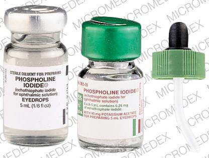


ECHOTHIOPHATE

History and source:

Eserine was isolated from Calabar beans. The alkaloid (physostigmine) was introduced as an anti-glaucoma agent in 1877 by Laqueur.

Official name:

Echothiophate is chemically Echothiophate iodide, 2-[(Diethoxyphosphinyl)-thio]-N,N,N-trimethylethanaminium iodide;2-mercaptoethyl) trimethylammonium iodide O,O-diethyl phosphorothiotae C9H23INO3PS.

Pharmacology:

This indirectly-acting parasympathomimetic agent is classified as a cholinesterase inhibitor or anticholinesterase. Cholinesterase inhibitors prolong the effect of Ach by inactivating the choliesterase enzymes which break it down.

Clinical pharmacology:

Echothiophate inactivates pseudocholinesterase and incompletely inactivates AChE, enhancing and prolonging the effects of AChE endogenously released from parasympathetic nerve endings.

Pharmaceutics:

A 0.125% solution is usually used.

Pharmacokinetics:

Onset of miosis is within 1 hour, IOP reduction occurs within 4 hours. Maximum miosis occurs within 2 hours and maximum IOP reduction within 24 hours. Miosis and IOP reduction can last for several weeks but usually lasts around 24-48 hours.

Therapeutic use:

Echothiophate is rarely used because of toxic effects. It was used in primary open-angle glaucoma, angle closure glaucoma after iridectomy and in accommodative esotropia.

Side effects and toxicity:

Side effects include corneal toxicity, conjunctival and intraocular vascular congestion, fibrinous iritis, individuals predisposed to retinal detachment, lacrimal canalicular stenosis, formation of posterior synechiae, iris cysts and cataracts.

Systemic toxic effects include diarrhea, nausea, abdominal cramps, general fatigue and weakness, hypotension and bradycardia.

Topical echothiophate makes patients more susceptible to prolonged paralysis following use of depolarizing muscle relaxants such as succinylcholine and procaine.

High risk groups:

Caution is advised in the following groups: Bronchial asthma, bradycardia, hypotension/hypertension, Down’s syndrome (echothiphate may cause hyperactivity), epilepsy, gastrointestinal disturbance, hyperthyroidism, iritis, myasthenia gravis, myocardial infarction, Parkinsonism, peptic ulcer, retinal detachment, urinary tract obstruction.


Tuesday, May 17, 2022

ANTI-FIBROSIS AGENTS IN GLAUCOMA


INTRODUCTION:

“Early” bleb failure after glaucoma filtering surgery (GFS) shows a hypercellular appearance, while “late” failure is associated with thicker collagen deposition.

Inhibition of scarring can be achieved at various levels by both physical and pharmacologic methods.

Decreasing the size of the operative site and careful hemostasis (which avoids excessive fibrin and thermal tissue injury) reduce the scope of the initial, intra-operative injury and subsequent fibrosis-stimulating inflammation.

Avoiding other sources of inflammation, such as combined surgical procedures, have the same effect. Successful egress of aqueous through the sclerostomy serves to keep the patency of the fistula for a prolonged time.

Characteristics of patients with increased risk for scarring include:

  • Young age
  • Black race
  • Previous unsuccessful GFS
  • Aphakia and pseudophakia
  • Neovascular glaucoma
  • Uveitic glaucoma
  • Iridocorneal endothelial syndrome
  • Previous prolonged topical glaucoma therapy
  • Conjunctival scarring from conditions such as alkali burns or pseudopemphigoid.


PHARMACOLOGIC AGENTS

Drugs used in inhibiting fibrosis in the GFS bleb can be categorized into the following groups=

1.        ANTI-INFLAMMATORY AGENTS

Corticosteroids

Non-steroidal anti-inflammatory drugs


2.       ANTI-NEOPLASTIC AGENTS

5-Fluorouracil (5-FU)

Mitomycin C (MMC)

3.       OTHER ANTINEOPLASTIC AGENTS

Antibiotics

Pyrimidine analogs  

Alkaloids


4.       OTHER ANTIFIBROSIS AGENTS

Interferon-α, calcium ionophore A23187, β-aminoproprionitrile (BAPN), and D-penicillamine (DPA)


1.      ANTI-INFLAMMATORY AGENTS

(a) Corticosteroids:

Sugar, first noted the beneficial effect of topical corticosteroids in prevention of bleb scarring.

Systemic steroids did not have any additional effects.

Subconjunctival steroids have been used at the conclusion of the GFS procedure.

The presumed mechanism of the anti-fibrosis activity of corticosteroids in GFS is the inhibition of the inflammatory response, mediated via blockage of the lipo-oxygenase and cyclo-oxygenase pathways by direct inhibition of phospholipase A2.

This results in decreased capillary permeability, chemotaxis inhibition, and suppression of fibrin deposition.

Decreased fibroblast proliferation occurs at higher concentrations while a stimulatory effect may be seen at lower concentrations.

(b) Non-steroidal anti-inflammatory drugs:

NSAIDs, inhibitors of both lipoxygenase and cyclooxygenase pathways, have demonstrated suppression of human ocular fibroblast proliferation.

However, in clinical trials, topical post-operative flurbiprofen 0.03% after GFS resulted in higher rate of encapsulated blebs and higher final IOP.

2.     ANTI-NEOPLASTIC AGENTS

Many pharmacologic agents hinder the scarring process via antimetabolic activity, typically interfering with one or more phases of the cell replication cycle of fibroblasts.

(a) 5-Fluorouracil (5-FU)=

5-Fluororidine (5-FUR) and 5-FU are pyrimidine nucleotide analogs. Similar to the pyrimidine analogs, they owe their anti-neoplastic activity to small structural dissimilarities from endogenous pyrimidines.

These agents require metabolic conversion to nucleotides to exert cytotoxicity. Simultaneous catabolism can inactivate these drugs.

5-FU is a fluorinated pyrimidine with a molecular weight of 130.08.

It undergoes intracellular conversion to the active deoxynucleotide, 5-fluro-2’-deoxyuridine 5’-monophosphate (FdUMP).

FdUMP causes competitive inhibition of thymidylate synthetase in the S-phase of the cell-replication cycle. This hampers the conversion of deoxyuridylic acid to thymidylic acid, thus impeding DNA synthesis.

FdUMP is also incorporated directly into DNA molecules after conversion by intracellular kinases to a triphosphate. Such DNA, with fluorouracil substituted for thymine, may be more unstable than native DNA.

It also interferes with RNA processing and function after its conversion to the ribonucleotide, fluouridine monophosphate (FUMP).

In GFS, this agent has been used mostly as post-operative subconjunctival injections.

The usual single dose is 5 mg (0.1 ml of undiluted bolus at 50 mg/ml).

The subconjunctival injection is performed 90 to 180 degrees away from the bleb. A tuberculin syringe with 30-gauge needle is used by going tangentially to the globe, bevel away from the sclera.

Avoid the conjunctival vessels to minimize bleeding.

Subconjunctival diffusion around the wound site may occur, especially through the needle track.

This elution of the drug into the tear film may encourage epithelial toxicity.

Leakage can be reduced by tamponade of the injection site with a cotton applicator; light massage to move the drug from the track and irrigation of residual undiluted 5-FU from the conjunctiva and eyelids with saline.

The number of injections can be adjusted by the clinical response and other factors such as patient access, acceptance, affordability, and compliance.

Intra-operative 5-FU implantation using a purified collagen sponge containing 100 µgm of 5-FU in the quadrant of surgery has also been reported. This leads to a slow release of the agent and potentially less epithelial toxicity.

Undiluted 5-FU (50 mg/ml) has been used intraoperatively using drug-soaked cellulose sponges placed under and over the scleral flap and sub-conjunctivally for a 5-minute period followed by copious balanced salt solution (BSS) irrigation.

Toxicity:

Systemic toxicity is rare as hardly 1-3% of the dose used in anti-cancer therapy enters the general circulation following ocular 5-FU usage.

Most of the drug is metabolized through hepatic clearance and elimination by respiration as carbon dioxide or renal excretion in the form of metabolites or free drug.

Ocular toxicity occurs as a consequence of its effect on rapidly dividing epithelial cells of the cornea and conjunctiva.

The Fluorouracil Filtering Surgery Study (FFSS) group reported punctate corneal epitheliopathy in 98% of patients, conjunctival epithelial defects, and corneal epithelial defects (64%). This toxicity was also responsible for a large number of conjunctival wound leaks.

Other corneal complications were: filamentary keratitis, keratinized corneal plaques, infectious corneal ulcers, and striate melanokeratosis (attributed to centripetal migration of pigment-laden stem cells from the limbus).

5-FU was also associated with punctal-canalicular stenosis, cicatricial ectropion due to lower lid dermatitis, contact dermatitis, and increased pigmentation of periocular skin.

The most devastating complications of 5-FU have been the occurrence of thin, cystic blebs, with an increased frequency of late bleb leaks, late endophthalmitis, and hypotonic maculopathy (also choroidal effusion and persistent shallow chambers).

5-FU should be avoided in patients with known corneal diseases that increase the risk of complications. Such conditions include bullous keratopathy, severe-dry eye syndrome, preexisting corneal epithelial defects, recurrent erosion syndrome, corneal melting syndrome, preexisting dellen, and conditions associated with decreased limbal stem cells such as Stevens-Johnson syndrome, pemphigoid, pseudopemphigoid, and old alkali burns.

Epithelial defects and wound leaks appear dose-related.

Careful closure of the conjunctival wound is critical to the prevention of early postoperative wound leaks.

Suturing with small taper point (round-bodied) needles with 8/0-10/0 sutures and employing a running mattress closure may prevent bleb leaks appreciably.

In general, antimetabolites are more commonly associated with hypotonic maculopathy in young and myopic patients.

The FFSS reported that 5-FU reduced the failure rates of GFS to 49% postoperatively, compared to 74% in control eyes.

(b) Mitomycin C (MMC)=

MMC was isolated by Wakaki and colleagues from Streptomyces caespitosus.

It undergoes enzyme activation in tissues and functions as an alkylating agent, cross-linking DNA.

Although it is cell-cycle phase non-specific, it is most active in the G and S phases of cell division.

MMC is used to treat neoplasms of the stomach, pancreas, bladder, colon, rectum, lung, cervix and breast.

In GFS it is used to prevent the replication of fibroblasts.

Studies have shown subconjunctival fibroblast proliferation inhibition to be dependent on the dose and exposure time.

The potency of MMC is 100 times greater than 5-FU.

MMC impedes the future replication of even those cells which are not synthesizing DNA at the time of exposure.

The markedly hypovascular blebs following the use of MMC are probably due to toxicity to vascular endothelium and contribute to the decreased scarring.

Toxic damage to the ciliary body and resulting decreased aqueous production could also contribute to the lowered IOP.

MMC has been used in a concentration between 0.1-0.5 mg/ml. it remains stable for 7 days at room temperature and for 14 days when refrigerated.

Non-preserved MMC should be used within 24 hours.

MMC is placed on the scleral bed after conjunctival dissection, either before or after the scleral flap has been fashioned. However, it should never be used once the coats of the eyeball have a full-thickness entry.

Some surgeons prefer to place the sponge over the scleral flap.

Care should be taken to avoid touching the edges of the conjunctival flap to the sponge, which might encourage postoperative wound leaks.


Toxicity:

MMC should be avoided during pregnancy due to its potential teratogenic effects.

It has significant corneal endothelial toxicity.

Scleral thinning and necrosis have not been reported following the use of MMC as described here.

MMC has fewer rates of corneal epithelial toxicity as compared to 5-FU.

Conjunctival wound leaks and hypotonic maculopathy is dependent on exposure times.

Treatment options for hypotonic maculopathy include autologous blood injection into the bleb, cryotherapy, and topical application of trichloracetic acid.

3.     OTHER ANTINEOPLASTIC AGENTS

 (a) Antibiotics=

Bleomycin, daunorubicin, doxorubicin, and mithramycin are antineoplastic antibodies extracted from Streptomyces. These agents have been found to inhibit fibroblasts.

(b) Pyrimidine analogs=

Cytosine arabinoside (Ara-C), trifluorothymidine, 5-fluoroorotate and metabolites of 5-FU have antiproliferative actions. They inhibit DNA polymerase and impede DNA synthesis.

(c)  Alkaloids=

Some alkaloids extracted from plants have been found to inhibit fibroblasts. These include vincristine, vinblastine and taxol.

4.     OTHER ANTIFIBROSIS AGENTS IN GLAUCOMA SURGERY

Interferon-α, calcium ionophore A23187, β-aminoproprionitrile (BAPN), and D-penicillamine (DPA) have been studied for their ability to inhibit cellular proliferation, collagen synthesis, and maturation.

Interferon-α inhibits collagen production, fibroblast proliferation, and chemotaxis.

Calcium ionophore A23187 also inhibits collagen synthesis.

DPA and BAPN inhibit collagen fibril cross-linking after synthesis, thus, decreasing the tensile strength of scar tissue.

Tissue plasminogen activators (TPA) is an enzyme that converts plasminogen to plasmin, which is fibrinolytic. TPA may enhance GFS.

Similarly, heparin also inhibits the proliferation of human scleral fibroblasts. However, it requires frequent exposure.


DEFERIPRONE IN GLAUCOMA MANAGEMENT

  Currently, lowering of intra-ocular pressure (IOP) remains the main therapeutic option for the treatment of glaucoma. However, studies hav...