Showing posts sorted by relevance for query CLASSIFICATION. Sort by date Show all posts

Wednesday, March 20, 2019

CLASSIFICATION OF GLAUCOMAS


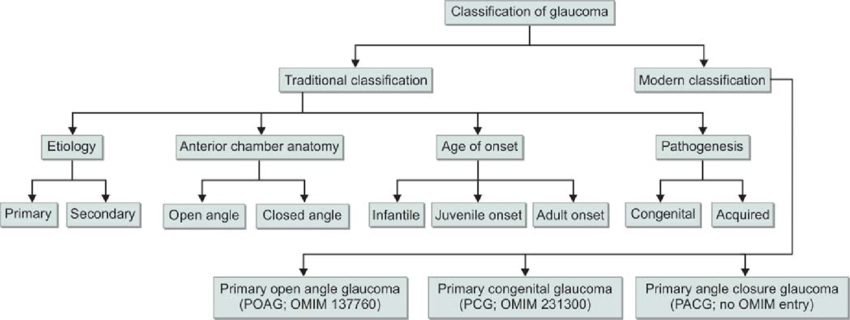
The classification of glaucomas has seen a progressive change as our understanding of this condition has evolved. The anatomic, gonioscopic, biochemical, molecular and genetic basis for the classification of glaucomas has been utilized, each having its own pros and cons. With the advent of new instruments to diagnose glaucoma, classifications have also been created based on the techniques utilized. Thus, the classifications of glaucoma include those based on:

-Etiology
-Mechanism
-Staging
-Optic nerve appearance
-Visual field damage
-Standard HRT parameters by bagging classification trees
-Automated classification of glaucoma stages using higher order cumulant features
-Texture features using neural networks

Unfortunately, none of the classifications have been satisfactory in their attributes to describe glaucoma. This is not unexpected, since there are different mechanisms of the disease and multifactorial pathogenetic factors at work in different individuals. Presently, the classification of glaucomas based on etiology and mechanism is still applied in clinical practice, having stood the test of time over the years.

Etiologic Classification= This is based on the underlying disorder causing alteration in aqueous inflow/outflow (Aqueous humor dynamics) or Retinal Ganglion Cell (RGC)/Optic nerve damage. 
Mechanistic Classification= This is based on specific alteration in the anterior chamber (AC) angle that causes intra-ocular pressure (IOP) to rise.

These classifications have incorrectly been based on our focus on elevated IOP as the major risk factor for the development of glaucoma, excluding other factors such as vascular, genetic or biochemical mechanisms among others.

CLASSIFICATION BASED ON ETIOLOGY 

Based on the etiology, glaucomas have been divided into primary and secondary. The primary glaucomas are assumed to have the initial events leading to outflow obstruction and IOP elevation primarily in the AC angle or conventional outflow pathway. These glaucomas are not associated with known ocular or systemic disorders which could impede aqueous outflow. They are usually bilateral and probably have a genetic basis. From a therapeutic standpoint it is essential to differentiate open angle glaucoma from closed-angle glaucoma.

On the other hand, secondary glaucomas are regarded as such because of a “partial understanding of the underlying, predisposing ocular or systemic events” [Bruce Shields]. These are usually asymmetric or unilateral. While some may have a genetic basis, others are acquired. As the concepts regarding the underlying causes of the glaucomas continue to develop, the primary and secondary classifications have become increasingly artificial and inadequate.

Classification of childhood glaucomas, especially those associated with developmental anomalies of the anterior chamber angle have been dogged by overlapping and variably defined nomenclatures which frequently denotes the age of onset rather than the underlying mechanism for the glaucoma. 

Bruce Shields has recommended replacing traditional concepts with a new scheme that provides a “better working foundation for the concepts of mechanism, diagnosis and therapy that will shape the management of glaucomas for the foreseeable future”. He has classified glaucomas based on staging. According to him, glaucomas can be considered to consist of 5 stages:

Stage I: Initiating events
Stage II: Structural alterations
Stage III: Functional alterations
Stage IV: RGC and ON damage
Stage V: Visual loss


The initiating events (Stage I) are speculated to have a genetic basis. Structural changes may start occurring in the RGCs or optic nerve head (ONH), as a result of alterations in proteins in these regions. These structural alterations (Stage II) could be subtle tissue changes in the blood vessels supplying the ONH or in supportive elements of the lamina cribrosa. Or they could act through mechanisms as yet to be understood. Structural changes may lead to functional alterations (Stage III) such as reduced axonal conduction, vascular perfusion to axons in the ONH or a progressive deformity of the lamina cribrosa that may lead (alone or in conjunction with a relative IOP elevation) to glaucomatous optic neuropathy (Stage IV), which gets reflected in subsequent VF changes (Stage V).

Traditionally, glaucomas have been divided into open and closed angle.

Chronic open angle glaucoma: This is characterized by optic nerve damage in an eye which does not have evidence of angle closure on gonioscopy and there is no identifiable secondary cause. Apparently inherited susceptibilities lead to increased resistance to aqueous outflow and higher vulnerability of the ONH to the level of IOP.

Pupillary block glaucoma: Primary Angle Closure includes asymptomatic individuals with occludable angles who have not had an acute attack, as well as those who had an attack which resolved spontaneously or with treatment prior to the development of any detectable nerve damage. Primary Angle Closure Disease (PACD) has been classified by the International Society for Geographical and Epidemiological Ophthalmology (ISGEO) into:

(1) Primary Angle Closure Suspect (PACS): Such eyes have iridotrabecular contact for atleast 2700 and normal IOP, ONH and VFs.
(2) Primary Angle Closure (PAC): There is iridotrabecular contact for atleast 2700 and raised IOP and/or peripheral anterior synechiae (PAS), but with normal ONH and VFs.
(3) Primary Angle Closure Glaucoma (PACG): There is PAC with evidence of glaucomatous damage in the ONH or VFs.
(4) Acute Angle Closure Crisis: There is periocular or ocular pain, often accompanied by headache, nausea or vomiting, IOP >21 mmHg, circumcorneal congestion, corneal edema, mid-dilated pupil and shallow anterior chamber.

Developmental anomalies of AC Angles:
These represent incomplete development of structures in the conventional aqueous outflow pathway. These anomalies could be inherited or acquired during intra-uterine life and lead to elevation of IOP. In some cases the developmental anomaly is not associated with primary or systemic etiologies and regarded as primary.

Pediatric glaucomas have been classified into the following categories:

(1) Primary Congenital Glaucoma (PCG): Primary congenital glaucoma that occurs at or shortly after birth or glaucoma of any etiology that occurs in the same time frame.
(2) Primary Infantile Glaucoma: It is genetically identical to PCG but presents 1-2 months after birth.
(3) Juvenile Open Angle Glaucoma: There is no ocular enlargement; absent congenital ocular anomalies or syndromes; Open, normal appearing angles; meets the glaucoma definition.
(4) Developmental Glaucoma: This term has been used as a giant waste basket for nearly all childhood glaucomas that are not acquired immediately after birth.

Glaucomas associated with other ocular disorders
This class includes those glaucomas in which the initiating event is an abnormality of the ocular structures such as corneal endothelium, iris, ciliary body, lens, vitreous, retina and so on. Or the initiating event is a definite second ocular pathology such as tumor, hemorrhage, inflammation and so on. Secondary glaucomas are properly considered to represent those eyes in which a second form of ocular pathology has caused IOP to rise above the normal range with consequent ON damage. The second ocular pathological processes causing optic neuropathy may include=
      i.        Neovascularization
     ii.        Uveitic conditions
    iii.        Trauma
   iv.        Lens-related

CLASSIFICATION BASED ON MECHANISM

Elevated IOP is the major risk factor for the development of glaucoma. However, the concept that statistically raised IOP is a defining characteristic of glaucoma has been almost universally discarded. A disadvantage of this mechanistic system is that it ignores the causes unrelated to IOP. Also, many of the glaucomas have more than one mechanism of outflow obstruction at different times in the course of disease. As a result some of the glaucomas must be classified under more than one mechanistic heading. On the plus side, the advantage of this classification is that our understanding of the mechanisms of aqueous outflow obstruction is usually more complete than our knowledge of initiating events. An understanding of the mechanism that leads to aqueous outflow obstruction is important in developing a rationale for controlling the IOP in each form of glaucoma.


Mechanisms of Open Angle Glaucoma=
The elements obstructing aqueous outflow may be located on the anterior chamber side of the trabecular meshwork [TM] (pretrabecular mechanisms); in the TM (trabecular mechanisms) or distal to the meshwork, in the Schlemm’s canal or further along the aqueous drainage system (post trabecular mechanisms).

Angle closure glaucoma mechanisms=
Angle closure mechanisms are the ones which cause apposition of the peripheral iris to the TM or peripheral cornea. The peripheral iris may be pulled (anterior mechanisms) or pushed (posterior mechanisms) into this position. In anterior mechanisms usually a contracting membrane in front of the iris pulls the iris towards the TM/peripheral cornea. It can also be caused by consolidation of inflammatory products in this area.
In posterior mechanisms pressure behind the iris, lens or vitreous causes the peripheral iris to be pushed into the anterior chamber angle. These mechanisms can occur with or without pupillary block. Pupillary block variants include pupillary block glaucoma in which there is apposition of the mid-periphery of the iris and the lens, thus blocking the egress of aqueous anteriorly through the pupil. The peripheral iris balloons in the form of “iris bombe”. The functional apposition in these patients is due to a genetically influenced configuration of the anterior segment of the eye. Such appositions may also be seen in lens-induced mechanisms such as phacomorphic glaucoma or ectopia lentis. Pupillary block can also occur from posterior synechiae. The “pushing” mechanisms can also occur without pupillary block such as ciliary block (malignant glaucoma), lens induced, forward shift of vitreous following lens removal, intraocular tumors, cysts of uveal tract, retrolenticular tissue contraction as in retinopathy of prematurity or persistent fetal vasculature.

Developmental anomalies of the AC Angles=
These represent incomplete development of structures in the conventional aqueous outflow pathway. Examples of these include: congenital glaucoma, Axenfeld-Reiger syndrome, Peter anomaly and iridocorneal adhesions.


Monday, July 21, 2025

COMPARISON OF DRI-OCT WITH HRT3


Glaucomatous optic neuropathy involves characteristic optic disc as well as retinal nerve fiber layer (RNFL) structural damage and related functional defects.

Tests of structural integrity include the Heidelberg Retinal Tomograph 3 (HRT3, Heidelberg Engineering GmbH, Heidelberg, Germany) and the continuously evolving technology of optical coherence tomography (OCT).


The HRT3 is a confocal scanning laser tomography (CSLO) device that uses a diode laser (670 nm) to scan the retinal surface at multiple consecutive parallel focal planes and produces repeatable and reproducible three-dimensional (3D) topographical images of the ONH and peripapillary RNFL. After image acquisition, the margins of the optic nerve head (ONH) need to be outlined by a manually drawn contour line to calculate ONH stereometric parameters. HRT3 also provides two different algorithms for ONH anatomy classification: the Moorfields regression analysis (MRA) that requires a contour line to be placed, and the newer contour-line independent Glaucoma Probability Score (GPS). The quantitative and objective measures of these structures are consequently classified as within normal limits (WNL), borderline, or outside normal limits (ONL) by automatic comparison with an ethnic-selectable normative database of eyes.


Deep range imaging OCT (DRI-OCT, Triton, Topcon, Tokyo, Japan) is a recently introduced swept-source OCT (SS-OCT) that uses a center wavelength of 1,050 nm and a bandwidth of approximately 100 nm compared to the fixed 850 nm wavelength of spectral-domain OCT (SD-OCT). The instrument achieves a high scan speed (100,000 A-scans/second) that allows for the acquisition of high-quality wide-field images containing both the ONH and the macula in a 12 mm × 9 mm single scan. SS-OCT, similar to SD-OCT, also provides separate standard macula and optic disc scan modes. Both thickness measurement values and normative comparisons are provided for all SS-OCT measurements.


DRI-OCT Triton: 3D wide(H) glaucoma report. A 75-year-old female with primary open-angle glaucoma in her left eye.
(A) Conventional color photography of the ONH. (B, C) macular GC analysis*. (D) Color-coded RNFL thickness map that corresponds to numeric RNFL thickness measurements. (E) SuperPixel-200 map. The uncolored pixels indicate the normal range, whereas the yellow- and red-colored pixels indicate abnormality at P = 1-5% and P < 1% of the normal level, respectively. (F) cpRNFL analysis*. (G) Numeric measurements of five ONH parameters


Kourkoutas and colleagues from Greece, have performed a study to determine the diagnostic performance of the ONH, macular, and circumpapillary retinal nerve fiber layer (cpRNFL) thickness measurements of wide-field maps (12 × 9 mm) using SS-OCT compared to measurements of the ONH and RNFL parameters measured by HRT3. 


They also evaluated the diagnostic ability of wide-field DRI-OCT thickness measurements (optic disc, RNFL, and macular) to differentiate glaucomatous from healthy eyes and compared them with the six main ONH stereometric parameters as well as with the GPS and MRA classification algorithms of the HRT3.


The authors found the highest sensitivities were achieved by the DRI-OCT categorical parameters of Superpixel-200 map and cpRNFL (12 sectors) thickness analysis. The best performing HRT3 continuous parameter was rim volume (AUC = 0.829, 95% confidence interval (CI) = 0.735-0.922), and the best continuous parameter for DRI-OCT wide-field was vertical CDR (AUC = 0.883, 95% CI = 0.805-0.951), followed by total cpRNFL thickness (AUC = 0.862, 95% CI = 0.774-0.951). Area under the curve (AUC) for disc area, rim area, linear CDR, and RNFL thickness were not significantly different between the two technologies. Using either the most or the least specific criteria, SuperPixel-200 map always showed the highest sensitivity among the categorical parameters of both technologies (82.1% and 89.7%, respectively). The highest sensitivity among HRT3 classification parameters was shown by MRA and GPS classification algorithms.


The study concluded that both wide-field DRI-OCT maps and HRT3 have good diagnostic performance in discriminating glaucoma from healthy eyes. However, DRI-OCT thickness values and normative diagnostic classification report the best performance.


REFERENCE:

Kourkoutas D, Triantafyllopoulos G, Georgiou I, Karamaounas A, Karamaounas N, Sotiropulos K, Kapralos D. Comparison of Diagnostic Ability Between Wide-Field Swept-Source Optical Coherence Tomography Imaging Maps and Heidelberg Retina Tomograph 3 Optic Nerve Head Assessment to Discriminate Glaucomatous and Non-glaucomatous Eyes. Cureus. 2022 Aug 19;14(8):e28188. doi: 10.7759/cureus.28188. PMID: 36158420; PMCID: PMC9482818.

Sunday, April 14, 2019

Area Under the ROC curve (AUC & ROC)


Area under the curve and Receiver operating characteristic are terms often used in studies and mentioned in articles, many of them related to glaucoma. However, for many of us some of these terms are abstract and appear to give no clue as to what they mean. This post takes a look at these terms: “Area under the curve” (AUC) and “Receiver operating characteristics” (ROC). Sometimes a combination term is used:”Area under the ROC curve” (AROC).

AUC ROC is one of the most important evaluation metrics for any classification model’s performance.

An ROC curve is a graph showing the performance of a classification model at all classification thresholds. This curve plots two parameters:
  • True Positive Rate (TPR)
  • False Positive Rate (FPR)

AUC stands for "Area under the ROC Curve." That is, AUC measures the entire two-dimensional area underneath the entire ROC curve.


What is ROC?:


Receiver Operating Characteristic (ROC) is a proven yardstick to measure the accuracy of diagnostic tests. The test divides the study population into positive (diseased) or negative (non-diseased). This is done by finding a cut-off or threshold which differentiates between diseased and non-diseased (e.g. IOP= 21 mmHg). The ROC Curve tells us about how good the model can distinguish between the two conditions. A good model can accurately distinguish between the two. Conversely, a poor model will have difficulty in separating the 2 test parameters.

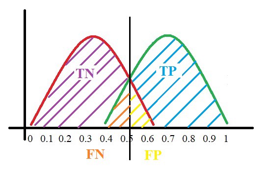
Let us assume we have a model which predicts whether the patient has a particular disease or not. The model predicts probabilities for each patient (in python researchers use the "predict_proba” function). Using these probabilities, we plot the distribution as shown below:


Here, the red distribution represents all the patients who do not have the disease and the green distribution represents all the patients who have the disease.


Now we pick a value where we need to set the cut-off i.e. a threshold value, above which we will predict everyone as positive (with disease) and below which will predict as negative (without disease). We will set the threshold at “0.5” as shown below:


All the positive values above the threshold will be “True Positives” and the negative values above the threshold will be “False Positives” as they are predicted incorrectly as positives.


All the negative values below the threshold will be “True Negatives” and the positive values below the threshold will be “False Negative” as they are predicted incorrectly as negatives.

Here, we have a basic idea of the model predicting correct and incorrect values with respect to the set threshold or cut-off.

To plot ROC curve, instead of Specificity we use (1 — Specificity) and the graph will look something like this:


So now, when the sensitivity increases, (1 — specificity) will also increase. This curve is known as the ROC curve.

Area Under the Curve:

The AUC is the area under the ROC curve. This score gives us a good idea of how well the model performs.

Let us take a few examples:


As we see, the first model does quite a good job of distinguishing the positive and the negative values. Therefore, in that curve the AUC score is 0.9 as the area under the ROC curve is large.


If we take a look at the last model, the predictions are completely overlapping each other and we get the AUC score of 0.5. This means that the model is performing poorly and it’s predictions are almost random.

Specificity gives us the True Negative Rate (TNR) and (1 — Specificity) gives us the False Positive Rate (FPR).

So the sensitivity can be called as the “True Positive Rate” (TPR) and (1 — Specificity) can be called the “False Positive Rate” (FPR).

So now we are just looking at the positives. As we increase the threshold, we decrease the TPR as well as the FPR and when we decrease the threshold, we are increasing the TPR and FPR.

Thus, AUC ROC indicates how well the probabilities from the positive classes are separated from the negative classes.


Limitations:


Unfortunately, diagnostic tests such as ROC have some limitations, such as the test may have a different sensitivity or specificity for the disease at different stages (for e.g. the test may give a different diagnostic yield in early glaucoma compared to advanced glaucoma). It may also be affected by covariates in the studied population (e.g. age, sex, rural-urban and co-morbidities). In such a mixed population, a single “pooled” ROC is often used as an average for the performance of the test. Regression analysis have also been done to assess the influence of covariates on the ROC curves.


REFERENCES: 

https://medium.com/greyatom/lets-learn-about-auc-roc-curve-4a94b4d88152

Saturday, October 7, 2023

JAPAN GLAUCOMA SOCIETY: CLINICAL PRACTICE GUIDELINES FOR GLAUCOMA


The Japan Glaucoma Society published the Clinical Practice Guidelines for Glaucoma in 2003, as well as the subsequent revised editions in 2006, 2012, and 2017. The fifth edition has been released in February 2023. 


The guidelines are presented in the following chapters, a short summary of which is being posted here.

https://link.springer.com/article/10.1007/s10384-022-00970-9

Chapter 1: Definition of Glaucoma

Glaucoma is a disease characterized by functional and structural abnormalities of the eye, with characteristic changes in the optic nerve and visual field, wherein optic neuropathy can be alleviated or suppressed by sufficiently lowering intraocular pressure (IOP).

Chapter 2: Classification of Glaucoma

The definition of glaucomatous optic neuropathy (GON) is given as, damage to the optic nerve associated with glaucoma. 

The chapter deals with the classification of glaucomas according to various mechanisms.

Primary open-angle glaucoma (broad) is a disease concept that encompasses both “primary open-angle”, where IOP is higher than the normal range, and “normal-tension glaucoma”. In clinical practice, primary open-angle glaucoma (broad) is divided into high IOP (primary open-angle glaucoma) and normal IOP (normal-tension glaucoma) groups.

Ocular hypertension

Patients with IOP above the statistically defined upper limits of normal; however, without abnormalities in the optic nerve or visual field

Preperimetric glaucoma (PPG)

The term PPG refers to a condition in which there are abnormalities suggestive of glaucoma, such as glaucomatous optic nerve head and retinal nerve fiber defects on ophthalmoscopy and optical coherence tomography (OCT), but no visual field defects are seen on conventional automated static perimetry test.

1. Primary angle-closure glaucoma (PACG):

PACG is a disease in which elevated IOP results from (primary) angle closure induced by genetic background or age-related changes in anterior segment morphology, without other factors, and in which glaucomatous optic neuropathy has already occurred.

2. Primary angle-closure (PAC):

PAC is a condition in which primary angle-closure causes elevated IOP or peripheral anterior synechia (PAS) but does not cause glaucomatous optic neuropathy. The name and etiology of this condition according to the speed of onset are the same as those of PACG.

3. Primary angle-closure suspect (PACS):

PACS is a condition in which there is primary angle-closure but without elevated IOP, organic PAS, or GON. Conversely, only appositional angle closure is present.

Some forms of PACG and PAC develop acutely and are collectively referred to as acute glaucoma attacks. In acute PACG and acute PAC, elevated IOP is often markedly high (40–80 mmHg), and symptoms such as decreased visual acuity, blurring of vision, glaucomatous halo, ocular pain, headache, nausea, vomiting, and diminished or absent light reflexes are common.

Secondary glaucoma is a condition in which elevated IOP is caused by other ocular diseases, systemic diseases, or drug use. Secondary glaucoma is classified according to the mechanism of elevated IOP.

The term childhood glaucoma refers to glaucoma resulting from a condition that develops in childhood. Although the term developmental glaucoma was used in the previous guidelines, the definition and classification have been substantially changed based on the recommendations of the World Glaucoma Association Consensus Conference. However, the upper age limit for childhood glaucoma has not been clearly defined by international standards.

Secondary childhood glaucoma is classified into glaucoma associated with non-acquired ocular anomalies and glaucoma associated with non-acquired systemic disease or syndrome. Those caused by acquired factors such as trauma, steroids, uveitis, and retinopathy of prematurity are classified as glaucoma associated with acquired conditions. Additionally, glaucoma that develops after cataract surgery, which is more frequent among acquired factors, is classified separately as glaucoma following cataract surgery.


Chapter 3: deals with the examination for glaucoma. It consists of the following parts:

i.                    Initial medical interview.

ii.                  Slit-lamp examination.

iii.                Assessment of IOP

iv.                 Gonioscopy

v.                   Fundus examination

vi.                 Visual field analysis


Chapter 4: is regarding treatment. It consists of the following parts:

i.                    Principles of glaucoma treatment

ii.                  Treatment practice


Chapter 5 is regarding glaucoma drugs.

Chapter 6 informs about laser procedures and their parameters.

Chapter 7 is regarding incisional surgery.

Chapter 8 deals with glaucoma treatment by disease type.


Thursday, March 12, 2020

WHAT IS GLAUCOMA?

(FOR PATIENTS)


INTRODUCTION

  • The term glaucoma is derived from the Greek word “glaukos” which means a bluish or greenish color of the pupil.


  • In the 10th century an Arab scholar, Ibn-Tabarri was the first to suggest that the eyes of glaucoma patients are harder than normal eyes.
  • Richard Bannister (1622) gave the concept of “Glaucoma Triad”. This triad consists of: Raised intra-ocular pressure (IOP), optic disc changes (cupping) and visual field defects.
  • The normal range of IOP is: 10-21 mmHg.
  • IOP is maintained as a balance between aqueous formation and aqueous outflow. This is called “aqueous humor dynamics”.


  • Later it was found that glaucoma could occur in the presence of normal levels of IOP. This is called “Normal Tension Glaucoma” (NTG).
  • Conversely, high IOP can be present without any glaucomatous changes. This is known as “Ocular Hypertension” (OHT) or Glaucoma Suspects.
  • Therefore, IOP is not the main criteria to define glaucoma.
  •  Glaucoma is now defined as: A multifactorial neurodegenerative disorder.

  • Glaucoma is the 2nd leading cause of blindness world-wide.
PATHOGENESIS
  • There are many theories regarding the pathogenesis of glaucoma.
  • These include: Mechanical, vascular, biochemical, genetic and other theories.
  • The mechanical theory explains the development of glaucoma by mechanical compression of the optic nerve head due to raised IOP.
  • However, it fails to explain the development of NTG and progression of glaucoma in cases where IOP is normal.
  •  The vascular theory explains glaucoma on the basis of deranged circulatory physiology and ischemia.
  • Certain biochemical molecules have also been implicated in the development of glaucoma. These include: glycine, aspartate, glutamate and others.
  •  Genetic linkage studies have revealed certain genes which have a very strong effect on disease causation. These include the MYOC and OPTN genes for familial POAG and CYP1B1 for congenital glaucoma.
TYPES OF GLAUCOMA
  • There are several systems available to classify glaucomas.
  • It can be based on etiology, where it defines the underlying disorder leading to alteration in aqueous humor dynamics or retinal ganglion cell (RGC) loss.
  •  Classification based on etiology can categorize glaucoma into “primary” or “secondary” forms. 
  • Primary glaucomas are those where the initial events leading to outflow obstruction and IOP elevation are confined to the anterior chamber angle or conventional outflow pathways with no apparent contribution from other ocular or systemic disorders.

Classification of glaucomas
OPEN ANGLE
DEVELOPMENTAL
ANGLE CLOSURE
Normal Tension
(NTG)
Primary (POAG)
Secondary
Primary
Secondary
Primary (PACG)
Secondary


Exfoliative Gl.

Pigment dispersion Gl.
Poorly formed or incomplete regression of TM.
Secondary to congenital ocular abnormalities.

Phacomorphic Gl.

Posterior segment tumors pushing the lens-iris diaphragm anteriorly.


INVESTIGATIONS
  

  • TONOMETRY= It is the procedure to measure IOP.
  • OPHTHALMOSCOPY= It is the procedure to observe the optic disc for signs of glaucoma (cupping) and fundus for cause of glaucoma (neovascularization, pigment dispersion).
  •  PERIMETRY= It is the procedure to map and analyze the visual field.
  • GONIOSCOPY= It is the procedure to observe the anterior chamber angle (differentiate between open and closed angle; look for secondary causes e.g. Neovascularization and pigment dispersion).
  • PACHYMETRY= It is the procedure to measure the central corneal thickness (CCT). (IOP measured is erroneously low in thin CCT; patients with thin CCT have higher risk of glaucoma).
  • RETINAL NERVE FIBER LAYER (RNFL) ANALYSIS= It is the procedure utilizing optical-coherence-tomography (OCT) to analyze the RNFL which becomes thinner with glaucoma progression.

TREATMENT


There are different methods available to control IOP. (IOP is the main risk factor and only risk factor which can be controlled presently).

1. Medical/pharmacologic
2. Laser
3. Surgical
4. Implants (MIGS, GDD, setons etc.)
5. Cyclodestructive procedures


Medical management:


·  INCREASE THE AQUEOUS OUTFLOW THROUGH TRABECULAR MESHWORK or UNCONVENTIONAL PATHWAYS=


·         Beta-blockers= Timolol, Betoxolol.

·         Prostaglandin analogues= Latanoprost, travoprost

·         Carbonic anhydrase inhibitors= Systemic: Acetazolamide; Topical: Dorzolamide

·         Alpha2 agonists= Brimonidine


·         REDUCE VITREOUS VOLUME (SHRINK THE VITREOUS)=

·         Hyperosmotic agents (systemic)= Mannitol, glycerol


Lasers:

·         Open angle glaucoma= Trabeculoplasty

·         Closed angle= Peripheral iridotomy


Surgical:

·         Trabeculotomy and Goniotomy (For developmental glaucomas)

·         Trabeculectomy

·         Non-penetrating glaucoma surgery (NPGS)


Implants:

·         Minimally Invasive Glaucoma Surgery (MIGS) = Express Mini Shunt; Xen Gel implant

·       Glaucoma Drainage Devices (GDDs) = Ahmed Glaucoma Valve; Baerveldt Glaucoma Valve; Molteno Glaucoma Valve


Cyclodestructive procedures (To destroy ciliary body, therefore reduce aqueous production):

·         Cyclocryotherapy/ Cyclocryopexy

·         Laser cyclophotocoagulation


NERVE GROWTH FACTORS FOR TREATMENT OF GLAUCOMA

  Neurotrophin-based therapies, such as recombinant human nerve growth factor (rhNGF), are promising candidates for non-IOP based treatments...