Saturday, June 29, 2024

TRANSPALPEBRAL ELECTRICAL STIMULATION


Felix Gil-Carrasco and colleagues have performed a study to analyze the effect of non-invasive transpalpebral specific exogenous voltages on intraocular pressure (IOP) in a cohort of open-angle glaucoma patients.


The electrical stimuli applied consisted of 10 Hz, biphasic, nonrectangular current pulses (100 μA) delivered from an isolated constant current stimulator.

The study was conducted on 78 eyes of 46 patients. Out of these, 58 eyes had reached maximum tolerated medical treatment, while 20 eyes were treatment naïve.

The mean baseline IOP in the treated POAG group was 19.25 mmHg ± 4.71.

The baseline IOP in the treatment-naive group was 20.38 mmHg ± 3.28.

After 4 months of follow-up, the mean IOP in the treated group was 14.41 mmHg ± 2.06 (P < 0.0001) (25.14% IOP reduction from baseline).

The mean IOP measurement in the treatment-naive group was 15.29 mmHg ± 2.28 (P < 0.0001) (25.9% IOP reduction from baseline).

The authors concluded that the IOP-lowering effect of transpalpebral electrical stimulation was statistically significant in the treated and treatment-naïve group of patients.

The authors claim that alterations in the mechanical properties of trabecular cells due to ion-channel dysfunction alter the volume and density of these cells. These changes subsequently have an adverse effect on the permeability and aqueous outflow through the trabecular meshwork.

The effect of transpalpebral-specific exogenous voltages is similar to tyrosine kinase inhibitors. The electric potential induces the reactivation of BKCa2+ in trabecular meshwork cells. This improves aqueous outflow through the conventional pathway. Since IOP is reduced in patients who have achieved maximal tolerable medical treatment, it is assumed that the aqueous outflow is improved through the non-conventional outflow pathway, and aqueous production is also reduced.

REFERENCE:

Gil-Carrasco F, Ochoa-Contreras D, Torres MA, Santiago-Amaya J, Pérez-Tovar FW, Gonzalez-Salinas R, Nino-de-Rivera L. Transpalpebral Electrical Stimulation as a Novel Therapeutic Approach to Decrease Intraocular Pressure for Open-Angle Glaucoma: A Pilot Study. J Ophthalmol. 2018 Jul 19;2018:2930519. doi: 10.1155/2018/2930519. PMID: 30116627; PMCID: PMC6079529.


Tuesday, June 25, 2024

ATHEROSCLEROSIS & GLAUCOMA RISK


Atherosclerosis is the thickening or hardening of the arteries caused by a buildup of plaque in the inner lining of an artery. The plaque comprises deposits of fatty substances, cholesterol, cellular waste products, calcium, and fibrin. As it builds up in the arteries, the artery walls become thickened and stiff affecting blood flow and compromising tissue health.


A study found carbonic anhydrase I (CA1), a CA family member, was highly expressed in atherosclerotic tissues of the aorta and stimulated atherosclerosis (AS) by promoting calcification. On the other hand, Methazolamide (MTZ), an oral CA inhibitor used in the treatment of glaucoma showed therapeutic and preventive effects on AS.

These findings suggest a relationship between AS and glaucoma.

A study by Song and colleagues in China was performed to confirm the validity of this hypothesis.

Among 152,425 AS patients, 1245 patients were also diagnosed with glaucoma (8.17‰). The incidence of glaucoma in the general hypertensive population (excluding AS) was 5.75% (1149/199,782 patients). The incidence of glaucoma in the AS patients was significantly higher (especially in those above 65 years of age) than in the hypertensive population and in the general population (both p values between groups were below 0.001).

In comparison, the incidence of glaucoma in patients without hypertension or AS was 1.79‰ (9513/5,313,433 patients).

Subgroup analysis showed that AS patients (especially those with arterial calcification) were more likely to suffer from angle-closure glaucoma (81.8%) than from open-angle glaucoma (18.2%).

MTZ decreased LDL levels while treating glaucoma. On the other hand, 5 patients (36%) had increased HDL levels after MTZ treatment. The average HDL level before treatment was 1.22±0.28 mmol/L, and the average value after MTZ treatment was1.46±0.37 mmol/L. This observation indicated that MTZ seemed to increase HDL levels following MTZ treatment for glaucoma, although the difference was not significant (p=0.08).

It can be surmised that the malfunction of CAs or the abnormal expression of their genes leads to the pathogenesis of various diseases, which could be interlinked.

REFERENCE:

Song, X., Li, P., Li, Y. et al. Strong association of glaucoma with atherosclerosis. Sci Rep 11, 8792 (2021). https://doi.org/10.1038/s41598-021-88322-4.


Friday, June 21, 2024

FAIRNESS CREAMS AND GLAUCOMA RISK


DR. JUWAIRIYA ILYAS

P.G. SCHOLAR, DEPARTMENT OF DERMATOLOGY,

NATIONAL INSTITUTE OF UNANI MEDICINE,

BENGULURU, INDIA

Skin lightening (SL) creams are currently big business world-wide. The advent of social media has brought physical beauty in increased focus, leading to increased usage of products (usually bought over-the-counter) to lighten the skin color. This artificial modification of the appearance is leading to increased usage of cosmetic products. Studies have shown that SL creams are used by 75% women in Nigeria, 60% in Senegal, 30% in Ghana, and 40% in South Korea.

61% of the skincare market in India consists of SL products. A study found 60% people using SL agents at least once in North India, and a similar number are reported from Malaysia. Similarly, 43.3% of Saudi women, and more than 60% of Jordanians reportedly use SL products. Dr. Mônica Manela-Azulay, Assistant Professor of Dermatology at the Santa Casa de Misericórdia of Rio de Janeiro, Brazil, mentioned that approximately 50% of all patients presenting to the clinic request topical treatments or procedures to lighten the skin.

In most cases patients are unaware that these products contain harmful substances such as steroids which can cause serious side-effects such as intractable glaucoma. [1, 2, 3]

Pollock et al. have performed a large International collaborative study and review of  a public health issue involving SL products. The objective of the study was to promote healthy skin and inclusive concepts of beauty in patients and society. According to the authors the risk of adverse reactions is increased when these products are used for prolonged periods of time or under occlusion. In some countries women smear creams on their bodies and wrap in dressings underneath their clothing. Individuals also may cover their skin with tight compression materials after applying creams or bathe in mixtures containing a combination of steroid agents, hydroquinone, bleach, and hydrogen peroxide. These individuals are at risk of local side effects on the skin, and also due to systemic absorption, which includes glaucoma.[4]

Dr. Neha Gupta from the famous L. V. Prasad Eye Institute in India has reported three cases of intractable glaucoma following use of fairness creams. The first case was a 19-year-old girl who reported using Pearly white skin whitening cream for 4–5 months prior to diagnosis of advanced open-angle glaucoma in both eyes. She had baseline intraocular pressures (IOP) 48- and 40-mm Hg in her right- and left-eye, respectively. As the pressures were uncontrolled with medical therapy, she underwent trabeculectomy in both eyes. The second case was a 21-year-old male who’s IOP was 42 mmHg in both eyes on presentation. He reported using Faiza skin whitening cream for 2 years. Subsequently, IOP was controlled medically. The third patient was a 29-year-old male patient who was using Pearly white (skin whitening) cream for 4 years. He underwent trabeculectomy in the left eye due to uncontrolled IOP.[5]

A Nigerian ophthalmologist, Dr Tarela Sarimiye, has been quoted as saying that indiscriminate and prolonged use of medications that contain steroids, including skin lightening creams, can affect the eyes, leading to glaucoma, the second leading cause of blindness in Nigeria.[6]

Use of steroid or bleaching creams by humans for beautification has been cited as one of the main causes of glaucoma in humans, Member of Parliament (MP) for Offinso North in the Ashanti region of Ghana, and ranking member on the Health Committee of Parliament, Collins Augustine Ntim, has stated. [7]

These reports and studies demonstrate the dark side of skin whitening or fairness creams and the necessity to regularly monitor individuals using them for glaucoma.


REFERENCES:

  1. Jose, A., Ray, J. G., & Schumacher, U. (2018). Toxic content of certain commercially available fairness creams in Indian market. Cogent Medicine5(1). https://doi.org/10.1080/2331205X.2018.1433104.
  2. Yusuf MA, Mahmoud ND, Rirash FR, Stoff BK, Liu Y, McMichael JR. Skin lightening practices, beliefs, and self-reported adverse effects among female health science students in Borama, Somaliland: A cross-sectional survey. Int J Womens Dermatol. 2019 Aug 23;5(5):349-355. doi: 10.1016/j.ijwd.2019.08.006. PMID: 31909156; PMCID: PMC6938842.
  3. JC Street, K Gaska, KM Lewis, ML Wilson. Skin bleaching: A neglected form of injury and threat to global skin.  African Safety Promotion Journal, Vol. 12, No. 1, 2014
  4. Pollock S, Taylor S, Oyerinde O, Nurmohamed S, Dlova N, Sarkar R, Galadari H, Manela-Azulay M, Chung HS, Handog E, Kourosh AS. The dark side of skin lightening: An international collaboration and review of a public health issue affecting dermatology. Int J Womens Dermatol. 2020 Sep 17;7(2):158-164. doi: 10.1016/j.ijwd.2020.09.006. PMID: 33937483; PMCID: PMC8072511.
  5. Gupta, Neha; Senthil, Sirisha. Intractable steroid-induced glaucoma due to skin whitening creams. Indian Journal of Ophthalmology - Case Reports 1(4):p 660-661, Oct–Dec 2021. | DOI: 10.4103/ijo.IJO_3757_20
  6. https://tribuneonlineng.com/skin-lightening-creams-linked-with-glaucoma/
  7. https://www.peacefmonline.com/pages/local/social/201403/192575.php


Tuesday, June 18, 2024

ASSOCIATION OF DERMATOLOGICAL CONDITIONS WITH GLAUCOMA


DR. YUSRA TANVEER

P.G. SCHOLAR, DEPT OF DERMATOLOGY

AJMAL KHAN TIBBIYA COLLEGE AND HOSPITAL

ALIGARH MUSLIM UNIVERSITY, ALIGARH, INDIA


Some types of glaucoma such as pseudo-exfoliative glaucoma (PXG) resemble certain integumentary disorders. In PXG a dandruff-like material is seen covering the ocular surfaces. The exact composition and pathogenesis of this pseudo-exfoliative material is unknown. However, the resemblance of this material to dandruff makes us wonder if glaucoma could be associated with dermatological conditions.

There appears to be a lack of studies in the literature regarding this association. Kang et al. reported a survey of 79,102 American women and 41,205 American men older than 40 years of age and at risk for glaucoma. They also reviewed the participants’ histories of skin cancer such as basal cell carcinoma (BCC), squamous cell carcinoma (SCC), and non-melanoma skin cancer. They found a 40% higher risk of PXG associated with a history of any non-melanoma skin cancer. This association was observed even with 4- and 8-year lags in non-melanoma skin cancer history. Also, the non-melanoma skin cancer association was stronger in patients younger than 65 than those older than 65. They also found that those living in northern latitudes had a significantly stronger association than those residing in southern latitudes. [1]

Another study was conducted by Ahmadpour et al. at the Mashhad University of Medical Sciences, Iran, to compare the frequency of dermatological manifestations between patients with PXG and those with primary open-angle glaucoma (POAG). The study involved 21 patients from the PXG group and 26 patients from the POAG group.

All PXG patients and 24 (92.3%) POAG patients had at least one abnormal dermatological finding (P = 0.495). [2]

The most common dermatological manifestations in patients with PXG were seborrheic dermatitis, dry skin, and cherry angioma. Other dermatologic manifestations of these patients included idiopathic guttate, hypomelanosis on the trunk and limbs, xeroderma pigmentosome and severe aging, lentigo maligna, and cafe au lait macules. In patients with POAG, the most common dermatological findings were seborrheic dermatitis, cherry angioma, and dry skin. Other specific manifestations seen in these patients included androgenic alopecia, telogen effluvium due to acetazolamide, nail-biting, pityrosporum folliculitis, sebaceous hyperplasia, nail fungal infection, alopecia areata, photosensitivity, macular amyloidosis, lipoma, and venous lake.

The most common dermatological condition found in all glaucoma patients was seborrheic dermatitis which was seen in 57.1–61.5%individuals. 12 patients with PXG and 16 with POAG had seborrheic dermatitis (P = 0.775). One patient in the PXG group and two in the POAG group had seborrheic findings on both head and face.

Eleven patients in the PXG group and seven in the POAG group had dry skin manifestations, mainly in their extremities. Although the frequency of dry skin was almost twice in those with PXG compared to the POAG group, the difference failed to reach the level of statistical significance (P = 0.130). Lentigines were observed in five PXG patients, while none of the POAG patients had this finding (P = 0.013).

There was no significant difference in the frequency of nevus and angiomas between the two groups. Also, no association between gender or patients’ age and the frequency of dermatologic manifestations (P > 0.154 for all comparisons) was found.

The frequency of dermatologic findings did not correlate with visual acuity, intra-ocular pressure, or cup-disc ratio (CDR) (P > 0.142 for all comparisons).

It is not clear how glaucoma is associated with skin disorders. One hypothesis is that glaucoma patients because of their age, lower socioeconomic status, and poor vision are unable to take care of themselves, leading to an increased frequency of these skin disorders. On the contrary, dermatological conditions could contribute directly to glaucoma, such as that seen in PXG. Also, corticosteroids used to treat skin diseases can contribute to steroid-induced glaucoma.

STEROID-INDUCED GLAUCOMAhttps://ourgsc.blogspot.com/search?q=steroid

Therefore, it may be advantageous to assess glaucoma patients for any dermatological conditions especially if they are being planned for surgery as any infections could be a potential risk. Also, steroid-induced glaucomas may confound the diagnosis unless specifically asked for as the patients may be reluctant to reveal their dermatological conditions themselves.

REFERENCES:

  1. Kang JH, VoPham T, Laden F, Rosner BA, Wirostko B, Ritch R, Wiggs JL, Qureshi A, Nan H, Pasquale LR. Cohort Study of Nonmelanoma Skin Cancer and the Risk of Exfoliation Glaucoma. J Glaucoma. 2020 Jun;29(6):448-455. doi: 10.1097/IJG.0000000000001496. PMID: 32487970; PMCID: PMC7317065.
  2. Ahmadpour F, Nahidi Y, Daneshvar R. Dermatological Findings in Glaucoma Patients: Comparison Between Pseudoexfoliative and Primary Open-angle Glaucoma. J Ophthalmic Vis Res 2022;17:479–485.


Saturday, June 15, 2024

WATSON AND BARNETT’S MODIFIED TRABECULECTOMY PROCEDURE


Prof. Peter Gordon Watson (30 April 1930 – 31 January 2017) was a British ophthalmologist, professor, and researcher.


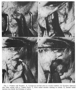
In an article published in the American Journal of Ophthalmology in 1975 Watson and Barnett presented a modification of Cairns’s trabeculectomy. [Watson PG, Barnett F. Effectiveness of trabeculectomy in glaucoma. Am J Ophthalmol. 1975 May;79(5):831-45. doi: 10.1016/0002-9394(75)90745-x. PMID: 1146946.]

Unlike Cairns’ so-called trabeculectomy procedure (which was more like a canaloplasty) in which aqueous was assumed to flow through the cut ends of the Schlemm’s canal, the Watson-Barnett modification consisted of excision of a block of sclero-cornea and allowing posterior filtration of aqueous through the sclerostomy (therefore, this procedure can be called a “sclerokeratectomy”).

The procedure was performed on 90 eyes in 60 patients between 1967 and 1972.

The follow-up ranged from one to six years postoperatively. (In 24 cases the follow-up was more than five years).

The procedure involved the creation of a wide conjunctival flap. This was followed with two radial incisions backward from the corneoscleral limbus, 4 to 5 mm long and 5 mm apart through two-thirds of the scleral thickness. The ends of these incisions joined a third circumferential incision in the sclera. This led to the formation of a scleral flap hinged anteriorly just into the clear cornea.

The deep flap containing the scleral spur and trabecular meshwork was now dissected. An incision through the remainder of the sclera was made transversely behind the scleral spur. It was readily identifiable by its fine white texture in contrast to the gray ciliary body posteriorly and the clear trabecular tissue with underlying brown iris tissue anteriorly.

Another pair of incisions were made passing through the full thickness of the cornea and sclera, and extending backward from the corneoscleral limbus posteriorly to the transverse incision.

The iris rarely bulged into the wound at this stage, but if it did, a small iridotomy released the aqueous humor from the posterior chamber and the iris fell back.

The superficial flap was replaced and sutured to the sclera. The number of sutures required depended on how well the incisions were apposed. If there was a retraction of the scleral edges, multiple or continuous sutures of 8-0 virgin silk or 10-0 Perlon or Ethilon were used. If the edges were well apposed, only three sutures of 8-0 virgin silk were needed in the posterior flap.

The conjunctiva was closed with continuous 8-0 virgin silk suture.


COMPLICATIONS:

INTRAOPERATIVE= In four eyes it was difficult to re-form the anterior chamber at the end of the procedure. One patient had prolonged bleeding from a ciliary process at the time of the iridectomy, however eventually it stopped spontaneously. The ciliary body prolapsed into the wound in another eye when the transverse incision was made in the deep flap.

POSTOPERATIVE= Flat AC was seen in two eyes on the first postoperative day and returned to normal later. Six eyes had shallow AC in the immediate few days after surgery and five eyes developed shallow AC later. Hyphemas large enough to form a fluid level were seen in 17 eyes (19%) postoperatively. Uveitis was seen in ten eyes. There was further loss of the visual field in one eye despite an average intraocular pressure of 15 mm Hg. In 16 eyes (18%) visual acuity deteriorated after operation. Progression of pre-existing cataract was seen in 14 eyes, corneal edema in one patient, and no cause was found in one patient.

RESULTS:

In 87% of the eyes, IOP was controlled without further medications or surgery. In 82 eyes (91%) a bleb formed, but in 8 (8.8%) the IOP was controlled with no evidence of bleb formation.

IOP was controlled at the outset in 84% of the eyes and eventually controlled in over 97%. Only 11% of the eyes required further medication and 5.5% further surgery.


Tuesday, June 11, 2024

CAIRNS' TRABECULECTOMY


The original objective of glaucoma surgery was to allow aqueous humor to exit more freely either through the sclera or into the suprachoroidal space. The former came to be called, generically, a glaucoma filtering procedure.


In his seminal paper published in 1968, John Edward Cairns described the goal of trabeculectomy as excising a short length of the Canal of Schlemm, with its trabecular adnexa, thus leaving two cut ends opening directly into the aqueous humor, with no trabecular tissue remaining as a barrier at that point, and restoring the integrity of the corneoscleral coat over the area of the excision. 


John Edward Cairns

Although naming this procedure trabeculectomy was appropriate (because the trabecular meshwork was removed to open Schlemm’s canal), the procedure might also have been accurately called canalostomy.

Cairns hoped that cutting open the edges of Schlemm’s canal would allow aqueous to leave without producing a filtering bleb; however, it became apparent that Cairns’ ‘‘trabeculectomy’’ only worked when a filtering bleb developed.

Pathology later showed fibrotic closure of the cut ends of Schlemm’s canal. Additionally, the presence of Schlemm’s canal in the trabeculectomy specimen did not correlate with outcomes.

The procedure that started as a ‘‘trabeculectomy’’ worked as a guarded filtration procedure, but the terminology remained unchanged.

Cairns reported 17 ‘‘trabeculectomy’’ procedures on eyes with uncontrolled glaucoma. After creating a flap starting in the cornea 2--3 mm anterior to the limbus and hinged on the sclera, a deep-scleral lamella containing Schlemm’s canal and trabeculum was excised and the flap was sutured firmly. A peripheral iridectomy was performed in seven patients. Seven of 17 patients developed a bleb, and histologic evaluation of the excised tissue in eight patients showed trabecular tissue.

Control of IOP for 10--14 weeks without subconjunctival drainage of aqueous humor occurred in about two-thirds of the patients. Cairns’ explanation for the reduction of IOP was the flow of aqueous through the open ends of Schlemm’s canal.

Further studies by Cairns and other investigators led to the conclusion that Cairns’ trabeculectomy was a ‘‘guarded filtering procedure’’.

Bleb formation occurred in most successful cases, histological studies on the excised block failed to show trabecular meshwork and patency of cut ends of Schlemm’s canal was observed in only a minority. In a few patients, the cut edges of Schlemm’s canal remained open. In these cases, compression of superficial scleral veins years after the surgery caused the passage of blood through the cut edges of Schlemm’s canal and into the anterior chamber. This finding indicated that at least some of Cairns’ trabeculectomies functioned as he intended.


Saturday, June 8, 2024

GLAUCOMA PREVALENCE IN INDIA


DR. SALEHA HASAN

MD (Community Medicine) PG resident,

Ajmal Khan Tibbiya College,

Aligarh Muslim University (AMU),

Aligarh, India


Community ophthalmology is a health-care modality promoting eye health and blindness prevention through initiatives that combine public health, community medicine, and holistic ophthalmic care approaches. Glaucoma is a significant cause of visual morbidity in India.

There are approximately 11.2 million persons aged 40 years and older with glaucoma in India.

The number of persons with Primary open-angle glaucoma (POAG) is estimated to be 6.48 (95% CI: 5.06-7.89) million.

Primary angle-closure glaucoma (PACG) affects an estimated 2.54 (95% CI: 1.88-4.28) million persons. Those with some evidence of damage—raised IOP, peripheral anterior synechiae or glaucomatous disc, or field changes comprise 6.62 (95% CI: 4.78-9.41) million persons. Those with any form of primary angle-closure disease could comprise 27.6 million persons. The prevalence of PACG and PAC show a linear increase with age.

The rates of ocular hypertension (OHT) show wide variations. Using the mean crude prevalence of 1.53% for OHT obtained from some studies conducted across India, an estimated 4.7 million persons could be ocular hypertensive.

Rates for secondary glaucoma are sketchily reported for the Indian population. They range from 0.08% to 1.39%. Crude estimates of those affected with secondary glaucoma, suggest around 2.28 million persons.

The proportion of those diagnosed to be bilaterally blind because of POAG in the APEDS, ACES, CGS (rural), CGS (Urban), and WBGS was 11.1%, 1.6%, 3.2%, 1.5%, and 5.2%, respectively. The corresponding figures for PACG for APEDS, CGS (rural), and CGS (Urban) were 16.6%, 2.9%, and 5.9%, respectively.

[Name key: Vellore Eye Survey (VES), Andhra Pradesh Eye Disease Study (APEDS), Aravind Comprehensive Eye Survey (ACES), Chennai Glaucoma Study (CGS), and West Bengal Glaucoma Study (WBGS).]

PACG on average produced 2 times the proportion of bilateral blindness than POAG.

These studies show that a community health approach to glaucoma is paramount in a country with a huge population, like India.

REFERENCE:

George R, Ve RS, Vijaya L. Glaucoma in India: estimated burden of disease. J Glaucoma. 2010 Aug;19(6):391-7. doi: 10.1097/IJG.0b013e3181c4ac5b. PMID: 20711029.


Tuesday, June 4, 2024

XEN-45 GEL IMPLANT: 2-YEAR OUTCOMES


A retrospective analysis of the prospectively collected data from the Fight Glaucoma Blindness observational registry was used to evaluate the real-world efficacy and safety outcomes of the Xen-45 gel stent implant over 24 months of follow-up.

SEE OTHER RELATED POSTShttps://ourgsc.blogspot.com/search?q=xen

Xen 45 was implanted in 646 eyes of 515 patients. [Xen stand-alone (n=199) and Xen+cataract (n=139)]

The most common diagnosis was primary open-angle glaucoma accounting for 69% of cases. Preoperatively, the mean IOP was 21.4±7.6mm Hg, the mean number of medications was 2.7±1.3 and the average visual field mean deviation where that was recorded, was −10.2±8.4dB.

Sub-group analysis included Xen implant alone versus Xen+Cataract surgery.

Complete success (CS) was defined as intraocular pressure (IOP) reduction ≥20% from preoperative and an IOP ≤18mm Hg and ≥6mm Hg with no secondary procedure at 2 years and without IOP-lowering medications.

Qualified success (QS) was defined similarly, allowing the use of IOP-lowering medications.

Preoperative IOP was 21.4±7.6 (mean±SD) mm Hg on 2.7±1.3 IOP-lowering medication and the mean deviation was −10.2±8.4 dB.

After 24-month follow-up, IOP was 16.8±7.3mm Hg (mean reduction of 21.7%) on 1.2±1.4 IOP-lowering medications, a mean reduction of 1.6 medications. The proportion of eyes which were completely off all glaucoma medications was 51%.

CS and QS rates at 24 months were 26% and 48%, respectively. CS and QS were higher in the Xen stand-alone group (33% and 52%, respectively) than in the Xen+cataract group (16% and 42%, respectively).

Success rates of 27% and 34% after Xen gel implant have been reported in other studies.

Bleb needling was performed in 28.4% of cases. Some other studies have reported rates of bleb needling following Xen implantation ranging from 41% to 62%.

18% of patients underwent a secondary glaucoma procedure, most commonly being bleb revision (7.5%). This included either bleb revision alone or revision combined with cataract surgery. In addition, 5.1% of eyes underwent trabeculectomy following Xen implantation within the first 24 months.

The most common adverse event was a loss of ≥10 letters of VA from baseline. This occurred in 31% of eyes within the first 6 months after surgery. This was attributed to ocular hypotony. Device exposure occurred in 1.8% eyes.

CONCLUSION:

The Xen 45 gel stent implant offers acceptable long-term efficacy for treating open-angle glaucoma. However, there is a significant rate of reoperation and needling, and outcomes are less effective if combined with cataract surgery.

REFERENCE:

Arnould L, Balsat E, Hashimoto Y, et al. Br J Ophthalmol Epub ahead of print: [please include Day Month Year]. doi:10.1136/ bjo-2023-325077.


Saturday, June 1, 2024

GINGER EXTRACT IMPROVES OCULAR BLOOD FLOW


It is known that glaucoma can develop or progress even in the presence of normal IOP. Deranged ocular blood flow (OBF) is suspected to be a major risk factor in such cases. Acute blood flow reduction in the retina contributes to axonal damage and retinal ganglion cell (RGC) death.


https://ourgsc.blogspot.com/search?q=blood+supply

Endothelin-1 (ET-1), one of the most potent vasoconstrictor peptides, has been implicated in glaucoma pathogenesis. Several studies have shown that intravitreal injection of ET-1 induces optic neuropathy in rodent models.

Takahashi and colleagues from Tohoku University Graduate School of Medicine have studied the effect of a ginger extract on optic nerve head blood flow (ONH BF) under ET-1 stimulation.

The researchers used laser speckle flowgraphy to study the ONH BF impairment profile under ET-1 stimulation. The main LSFG variable, expressed in arbitrary units, is termed mean blur rate (MBR) and indicates relative blood flow.

To establish the ONH BF impairment profile under ET-1 stimulation, brown Norway rats were administered an intravitreal injection of ET-1 under anesthesia. They were subsequently given ginger extract sublingually to assess its effect on ONH BF in both normal and ET-1-induced ischemic conditions.

The ginger extract contained 50% red ginger [Zingiber ofcinale var. rubra] extract and 50% cyclodextrin. The active ingredient in the ginger extract was [6]-shogaol. A previous study has shown that [6]-shogaol increases intestinal blood flow (IBF), and that this effect was mediated by the calcitonin gene-related peptide (CGRP) and its receptor.


The percentage changes in MBR in the case group were compared to the sham (PBS injection and non-intake). In the case (ET-1 injection and ginger extract intake) group, %MV was 64.7±19.2%. In the control (ET-1 injection and non-intake) group, %MV was 44.0±20.6%.

Percentage MBR in the vessels (%MV) and percentage MBR in the tissues (%MT) were significantly higher in the ginger extract intake group than in the non-ginger extract intake group (P=0.039, P=0.046, respectively), indicating that the ginger extract ameliorated the dysregulation of ONH BF. Similarly, the overall MBR (%MA) in the ginger extract intake group tended to be higher than the non-ginger extract intake group (P=0.068).

The study demonstrates that a ginger extract containing [6]-shogaol can increase ONH BF under conditions of good health as well as ischemia, independently of BP and IOP. The agent can act as a useful treatment modality for NTG and help prevent optic neuropathy induced by vascular dysregulation, such as that seen in NTG.

REFERENCE:

Takahashi N, Sato K, Kiyota N, Tsuda S, Murayama N, Nakazawa T. A ginger extract improves ocular blood flow in rats with endothelin-induced retinal blood flow dysfunction. Sci Rep. 2023 Dec 20;13(1):22715. doi: 10.1038/s41598-023-49598-w. PMID: 38123793; PMCID: PMC10733345.

A blind bat eating ginger

EXTREME WEATHER EVENTS AND GLAUCOMA

  Extreme weather events (EWEs) (e.g., heatwaves and cold spells) are reportedly associated with high morbidity through various physiologic...