Monday, September 30, 2024

NOVEL NDI1 GENE THERAPY


Several factors cause glaucoma, some working together in an individual. An emerging concept in the development of this disease is mitochondrial dysfunction and oxidative stress in the retinal ganglion cells (RGC), trabecular meshwork, and the lamina cribrosa (LC) in the optic nerve head.

Sophia Millington-Ward et al, have reported a study to assess the effectivity of an enhanced NDI1 gene (eNdi1) as a gene-independent gene therapy for glaucoma. The therapy is aimed at improving mitochondrial function and decreasing reactive oxygen species (ROS) in a variety of glaucoma models.


The study involved H2O2-insulted primary porcine LC cells (pLC cells). The control cells received eNdi1 constructs delivered via adeno-associated viral vectors (AAV-eNdi1).

H2O2 insult significantly reduced basal oxygen consumption rate (OCR) levels (70.6 ± 37.3% versus 100.0 ± 50.2%), maximal OCR levels (82.7 ± 23.3% versus 118.2 ± 28.3%), and ATP production (48.6 ± 32.4% versus 71.8 ± 37.7%), indicating that oxidative stress significantly reduces mitochondrial function in H2O2-insulted pLC cells.

Conversely, the basal OCR, maximal OCR, and ATP production levels were significantly higher in H2O2-insulted cells that had received AAV eNdi1 (123.9 ± 39.4%, 137.5 ± 32.3%, and 81.5 ± 28.1%, respectively), indicating rescue of mitochondrial function in the model.

 AAV-eNdi1-treated eyes displayed significantly improved photonegative responses compared to control PBS-injected eyes (6.4 ± 3.3 µV and 5.0 ± 3.1 µV, respectively, p < 0.05, paired t-test; n = 25).

eNdi1 directly increases mitochondrial function in cell models of glaucoma. Additionally, the study showed that ROS and mitochondrial stress were reduced, while cellular health was improved.

Therefore, directly increasing mitochondrial function is protective against the development of glaucoma and provides significant benefits.

REFERENCE:

Millington-Ward S, Palfi A, Shortall C, Finnegan LK, Bargroff E, Post IJM, Maguire J, Irnaten M, O Brien C, Kenna PF, Chadderton N, Farrar GJ. AAV-NDI1 Therapy Provides Significant Benefit to Murine and Cellular Models of Glaucoma. Int J Mol Sci. 2024 Aug 15;25(16):8876. doi: 10.3390/ijms25168876. PMID: 39201561; PMCID: PMC11354491.

Saturday, September 28, 2024

ALPHA-2 ADRENERGICS AND ALZHEIMER'S DISEASE


A study by Chou et al has shown that patients on topical alpha-2 adrenergic monotherapy have a significantly higher risk of developing Alzheimer’s disease (AD).

The nationwide population-based cohort study utilized insurance claims data from Taiwan’s National Health Insurance Research Database between 2008 and 2019. Participants were adults aged 45 years or older either with a diagnosis of glaucoma or without.

There were 202,000 participants included in the study, with 101,000 in each group (glaucoma and control groups).

Those with glaucoma received at least one antiglaucomatous medication (including α2-adrenergic agonists, cholinergic agonists, beta-blockers, prostaglandin analogs, and pilocarpine) for over 90 days. Those with pre-existing AD diagnoses before the index date were excluded.

Glaucoma patients on topical alpha-2 adrenergic agonist monotherapy exhibited a significantly higher AD risk (aHR 1.15, 95% CI=1.011.31) compared to those on beta-blockers.


Irrespective of the type of glaucoma, individuals with glaucoma had a significantly higher risk of AD compared to those without glaucoma (POAG: aHR 1.23, 95% CI=1.081.40; NTG: aHR 1.49, 95% CI=1.191.85; PACG: aHR 1.35, 95% CI=1.191.52; unspecified glaucoma: aHR 1.36, 95% CI=1.231.50).

The study concluded that topical alpha-2 adrenergic agonists might pose increased AD risk in individuals with glaucoma compared to beta-blockers. Therefore, they should be used cautiously, especially in middle-aged and older populations. The findings also suggest that glaucoma, regardless of the type may increase the risk of AD.

REFERENCE:

Chou, CC., Lu, YA., Weng, CH. et al. The association between antiglaucomatous agents and Alzheimer’s disease. Eye (2024). https://doi.org/10.1038/s41433-024-03348-y


Tuesday, September 24, 2024

METAMATERIAL ELECTRICALLY-PASSIVE SENSOR-EMBEDDED CONTACT LENS


The current treatment of glaucoma is limited to the control of intraocular (IOP). Conventionally, the IOP is measured only during the patient’s visit to the health-care worker. However, this approach has been found to have multiple limitations and does not provide adequate data to develop the target IOP. An emerging paradigm for the IOP measurement strategy is to record the circadian cycles of IOP variation over 24 hours and repeat this measurement periodically.

The continuous monitoring strategy has led to the development of many extra- and intra-ocular devices. Contact lens-based devices provide a practical approach to this method.

A metamaterial-inspired electrically-passive sensor-embedded contact lens for continuous intraocular pressure (IOP) monitoring has been developed by Kaya et al. The data is received by a wearable antenna patch. The sensing technology is based on the electrically passive sensor embedded in a disposable soft contact lens, and a wearable electronic readout system to collect, store, and process the data.


In comparison to the electrically active silicon-based sensors, this sensor is thin, flexible, and does not require power transmission.


A human study involving six healthy volunteers was performed under various experimental conditions and the system was found to be robust in its functions.

REFERENCE:

Kaya O, Akif Aydin M, Teymoori M, Kaan Erden O, Sadeghzadeh S, Dedeoglu UO, Demir S, Muhikanci O, Sahin A, Torun H, Dundar G, Yalcinkaya AD. A first-in-human pilot study of a novel electrically-passive metamaterial-inspired resonator-based ocular sensor embedded contact lens monitoring intraocular pressure fluctuations. Cont Lens Anterior Eye. 2024 Apr;47(2):102102. doi: 10.1016/j.clae.2023.102102. Epub 2023 Dec 19. PMID: 38114379.


Saturday, September 21, 2024

ENDOSCOPIC CYCLOPHOTOCOAGULATION


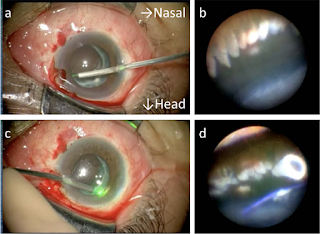
Endoscopic cyclophotocoagulation (ECP) is a procedure to lower aqueous production by laser-induced destruction of the ciliary processes, the site of aqueous production in the eyes. The key feature of ECP is direct visualization of the ciliary processes as the target tissue for controlled laser ablation. With this procedure, it is possible to titrate the extent of ciliary body ablation to maximize IOP lowering while minimizing collateral damage and adverse events. It can be performed along with cataract surgery or as a stand-alone treatment.


The ECP instrumentation consists of the laser endoscope and the console to which it is attached. The laser endoscopy console combines a 175 W xenon light source for illumination, 810 nm diode laser for photocoagulation, helium-neon laser aiming beam, and video imaging for intraocular visualization.


The endoscopy probe contains all three fiber groupings and is available in 19, 20, or 23 gauge sizes with a field of view ranging from 70° to 140° and depth of focus spanning 1–30 mm. The probe tips are straight or curved and easily fit through a 2.0 mm clear corneal incision. Another advantage to the 23-gauge probe is its compatibility with all 23-gauge vitrectomy trocar systems. The probes can be sterilized and reusable up to 25 times or more.

A variety of anesthesia may be used for ECP including intracameral, sub-Tenon's, or retrobulbar routes of administration. If intracameral anesthesia is utilized, increased intravenous sedation may be needed to maximize patient comfort during the laser application.

Before the start of the procedure, the three component cables of the ECP probe should be securely connected to the laser console. The camera image should be focused with the desired orientation and illumination adjusted outside the eye before the initiation of surgery. The laser should be set to continuous duration with an initial power of 0.25 W and an aiming beam setting of 20–30.

A temporal or superiorly placed clear corneal incision is performed near the limbus, approximately 2.0 mm in width. The ciliary sulcus is deepened with cohesive viscoelastic to improve visualization of the ciliary processes. The probe is then inserted through the corneal wound and positioned in the sulcus at or near the pupillary border.

The surgeon then directs his/her gaze towards the monitor to gain orientation in the sulcus and identify the target tissue. During treatment, approximately 6-7 ciliary processes should be in view at all times as this places the probe at an optimal distance for absorption of laser energy. Once the aiming beam is placed over a ciliary process, the foot pedal is depressed to deliver laser energy continuously. Treatment is titrated according to the visualized tissue response. The process should whiten and shrink to a variable degree after appropriate treatment.

If the probe is closer to the processes, a shorter duration and/or lower power will be needed to reach the desired effect. Rupture or popping of the processes should be avoided as an indication of over-treatment. The probe can then be advanced along the adjacent processes while applying laser energy. The entire visible area of each ciliary process should be treated including anterior and posterior edges as well as crypts in between processes.


Treatment should be carried to the extent of visualization in one direction, and then the probe is rotated 180° with rotation of the image on the monitor, and treatment is continued as far as possible in the other direction. With a curved probe, a single incision allows treatment of approximately 270° of ciliary processes. If more treatment is desired, a second incision may be placed 180° away from the initial wound to gain access to the subincisional processes and complete a 360° treatment for additional IOP lowering.

In aphakic or pseudophakic patients, a pars plana approach (ECP plus) may also be utilized to achieve a more thorough treatment of the ciliary processes when aggressive IOP lowering is desired. A standard 2 or 3 port pars plana vitrectomy must be performed initially, followed by insertion of the probe through one of the sclerotomies. Once the processes are visualized, treatment is carried out in the same fashion as the anterior approach. The anterior 1-2 mm of pars plana may also be treated in severe, refractory cases but may increase the risk for hypotony postoperatively.


At the end of the procedure viscoelastic is removed and the incision/s coapted.

Postoperatively the patient is started on steroid drops until the inflammation resolves. The anti-glaucoma medications are continued until the inflammation subsides and the desired IOP is achieved.

REFERENCE:

Seibold LK, SooHoo JR, Kahook MY. Endoscopic cyclophotocoagulation. Middle East Afr J Ophthalmol. 2015 Jan-Mar;22(1):18-24. doi: 10.4103/0974-9233.148344. PMID: 25624669; PMCID: PMC4302471.


Tuesday, September 17, 2024

MECHANICAL THEORY OF GLAUCOMA: ARTICLE PUBLISHED


The mechanical theory of glaucoma is one of the oldest theories regarding the development and progression of glaucoma. Initially, it was presented as a simple cause-and-effect mechanism. The high pressure compressed the lamina cribrosa in the optic nerve head, causing cupping and damage to the axons. However, now we know that multiple factors are working around the optic nerve head and they can collectively cause glaucomatous optic nerve degeneration. These factors can be studied in terms or pre-laminar, laminar, and post-laminar factors.


This article on these factors has been published in the current issue of the Taiwan Journal of Ophthalmology. For open access to the article kindly follow the link given here.

https://journals.lww.com/tjop/fulltext/2024/14030/the_mechanical_theory_of_glaucoma_in_terms_of.10.aspx


Saturday, September 14, 2024

365 BREATHING TECHNIQUE IN GLAUCOMA


DR. IRAM JAHAN

P.G SCHOLAR

DEPARTMENT OF AMRAZE AIN UZN ANF HALAQ WA ASNAN

STATE UNANI MEDICAL COLLEGE

PRAYAGRAJ, INDIA


A study has been performed, to assess the effect of the "365 breathing technique" on intraocular pressure (IOP), autonomic functions, and stress biomarkers in patients with primary open-angle glaucoma.

The randomized, controlled, interventional trial involved 40 patients in the intervention group who followed the "365 breathing" (three times a day, breathing rate: 6 cycles/min for 5 min) protocol in addition to their pharmacological glaucoma treatment. [SEE DETAILS BELOW]

The control group also had 40 patients who continued only with their pharmacological glaucoma treatment.

IOP, serum cortisol, heart rate variability (HRV), and heart rate response to deep breathing test (DBT) were recorded at preintervention and 6 weeks postintervention.

At baseline, the mean IOP, serum cortisol, parameters of the HRV test, and DBT were comparable between the two groups.

At the 6-week follow-up, in the intervention group, mean IOP was significantly lower (16.09 ± 2.24 vs 18.38 ± 1.58 mm Hg, P = 0.03) and serum cortisol was significantly lower, as well (13.20 ± 3.11 vs 14.95 ± 2.60 mcg/dl, P = 0.038) compared with the control group.

In the HRV test, analysis showed a higher postintervention parasympathetic activation in the intervention group. In the intervention group at 6 weeks (1.65 vs 1.79, P = 0.019), the sympathovagal balance shifted towards greater vagal modulation. There was a significant increase in delta heart rate (P = 0.019) and expiratory: inspiratory ratio (P = 0.011) in the intervention group at 6 weeks compared to the baseline values, indicating improved parasympathetic reactivity to DBT.

 CONCLUSION:

The "365 breathing" technique can reduce IOP and serum cortisol, and improve autonomic dysfunction in patients with glaucoma.

 REFERENCE:

Dada T, Gwal RS, Mahalingam K, Chandran DS, Angmo D, Gupta S, Velpandian T, Deepak KK. Effect of "365 Breathing Technique" on Intraocular Pressure and Autonomic Functions in Patients With Glaucoma: A Randomized Controlled Trial. J Glaucoma. 2024 Mar 1;33(3):149-154. Doi: 10.1097/IJG.0000000000002356. Epub 2024 Jan 9. PMID: 38194285.


Breathing Strategies for Stress Relief:

If you find that your day, or even moments throughout the day, are marked by stress, the following breathing strategies may be techniques to try. To enhance these breathing methods, sit or stand straight, relax your body, remove distractions, and focus on the sensation of your breath. When thoughts begin to distract you, put them aside and redirect your attention to your breath. If you begin to feel dizzy, resume your regular breathing pattern.

365 Method

  • Three times a day, breathe six breath cycles per minute for five minutes.
  • One breath cycle means to inhale for 5 seconds and exhale for 5 seconds.
  • This breathing method can bring immediate feelings of relaxation.
  • Practice this technique every day (365 days) for maximum benefit.

PLEASE FOLLOW THE LINK GIVEN BELOW FOR FURTHER INFORMATION ON BREATHING TECHNIQUES:

 https://extension.sdstate.edu/using-your-breath#:~:text=365%20Method,365%20days)%20for%20maximum%20benefit


Tuesday, September 10, 2024

STATIN USE AND GLAUCOMA


DR. MOHSINA

P.G. SCHOLAR

DEPARTMENT OF ANATOMY

STATE TAKMEEL-UT-TIB COLLEGE & HOSPITAL

LUCKNOW, INDIA


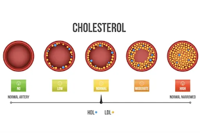
Cholesterol is the principal sterol of all higher animals, distributed in body tissues, especially the brain and spinal cord, and in animal fats and oils. Elevated cholesterol in the blood, especially when bound to low-density lipoprotein (LDL, often called "bad cholesterol"), may increase the risk of cardiovascular disease.


Statins are a group of medicines that reduce the production of low-density lipoprotein (LDL) cholesterol in the liver, thus lowering blood cholesterol levels.

Statins are usually used to lower blood cholesterol levels and reduce the risk for atherosclerosis-related illnesses, such as cardiovascular disease. 

Statins have been suggested to increase retinal and choroidal blood flow, protect retinal ganglion cells in the setting of ischemia, and lower IOP, and they have been investigated as neuroprotective agents outside their primary indication as anti-hypercholesterolemic agents.


A study was performed by Lee and associates to investigate the association between statin use and glaucoma in the 2017-2022 All of Us (AoU) Research Program of the National Institute of Health (NIH), USA.

The cross-sectional, population-based study involved 79,742 adult participants aged ≥ 40 years with hyperlipidemia enrolled in the AoU database.

Of the 79,742 individuals with hyperlipidemia in AoU, there were 6,365 (8.0%) statin users.

Statin use was associated with increased glaucoma prevalence when compared with statin non-use (adjusted odds ratio [aOR]: 1.13, 95% confidence interval [CI]: 1.01-1.26).

Higher serum levels of LDL were associated with increased odds of glaucoma (aOR: 1.003, 95% CI: 1.003, 1.004).

Statin users had significantly higher LDL levels in comparison to the nonusers (144.9 mg/dL versus 136.3 mg/dL, p-value < 0.001).

Analysis stratified by LDL identified positive associations between statin use and prevalence of glaucoma among those with optimal (aOR = 1.39, 95% CI = 1.05-1.82) and high (aOR = 1.37, 95% CI = 1.09-1.70) LDL levels.

Age-stratified analysis showed a positive association between statin use and the prevalence of glaucoma in individuals aged 60-69 years (aOR = 1.28, 95% CI = 1.05-1.56).

CONCLUSIONS:

The study concluded that statin use was associated with increased glaucoma likelihood in the overall adult AoU population with hyperlipidemia, in individuals with optimal or high LDL levels, and individuals 60-69 years old. Findings suggest that statin use may be an independent risk factor for glaucoma, which may furthermore be affected by one’s lipid profile and age.

REFERENCE:

Lee SY, Paul ME, Coleman AL, Kitayama K, Yu F, Pan D, Tseng VL. Associations between Statin Use and Glaucoma in the All of Us Research Program. Ophthalmol Glaucoma. 2024 Jul 31:S2589-4196(24)00137-6. doi: 10.1016/j.ogla.2024.07.008. Epub ahead of print. PMID: 39094953.


Saturday, September 7, 2024

Association of HYPERTENSION and GLAUCOMA


DR. SHIBRA FAROOQ

P.G. SCHOLAR

DEPARTMENT OF ILAJ-BIT-TADBEER

AJMAL KHAN TIBBIYA COLLEGE,

ALIGARH MUSLIM UNIVERSITY, INDIA


This post discusses two meta-analyses that analyze the association of hypertension with glaucoma.

META-ANALYSIS 1:

Bae HW, Lee N, Lee HS, Hong S, Seong GJ, Kim CY. Systemic hypertension as a risk factor for open-angle glaucoma: a meta-analysis of population-based studies. PLoS One. 2014 Sep 25;9(9):e108226. doi: 10.1371/journal.pone.0108226. PMID: 25254373; PMCID: PMC4177901.

The association between hypertension and glaucoma is controversial. It is usually assumed that hypertension increases the risk of development and progression of glaucoma.

In the Blue Mountains Eye Study, hypertension increased the risk of open-angle glaucoma (OAG) by more than 50%. The Egna-Neumarkt Study also found an association between OAG and hypertension. However, the Barbados Eye Study did not find an association between the two.

Hypertension can cause damage to the optic nerve through multiple mechanisms. These include direct microvascular damage, impairing the blood flow to the anterior optic nerve; auto-regulation of the posterior ciliary circulation; and induction of hypotensive episodes, especially at night, by anti-hypertensive therapy, which can injure the optic nerve.

A meta-analysis to look into systemic hypertension as a risk factor for OAG was performed by Bae et al. The meta-analysis included 16 studies involving 60,084 individuals.

The present meta-analysis of population-based studies showed that individuals with hypertension have an approximately 1.2-fold higher risk of developing OAG than individuals without hypertension. The pooled odds ratio (OR) was 1.22 (95% CI: 1.09–1.36) using the fixed-effects model and 1.22 (95% CI: 1.08–1.37) using the random-effects model.

The study found that hypertension increased the risk of OAG development irrespective of age.

The risk of developing high-tension glaucoma (HTG) was increased to a greater extent by hypertension than that of OAG, whereas the risk of developing normal-tension glaucoma (NTG) was not affected by hypertension (OR=1.92 and 0.94, respectively). This difference might be attributable to the difference in IOP, which is related to ocular perfusion pressure (OPP).

There are two conflicting mechanisms involving hypertension and glaucoma. Increased BP leads to reduced ocular blood flow due to thickening and stiffening of the vessel wall, thereby increasing the risk of OAG. The second mechanism is that a higher BP produces a higher OPP, which could decrease the risk of OAG.

No significant regional differences between Eastern and Western populations were noted.

META-ANALYSIS 2:

Nislawati R, Taufik Fadillah Zainal A, Ismail A, et al. Role of hypertension as a risk factor for open- angle glaucoma: a systematic review and meta-analysis. BMJ Open Ophthalmology 2021;6:e000798. doi:10.1136/ bmjophth-2021-000798.

The relationship between blood pressure (BP) and the incidence of glaucoma is still being debated.

Several studies have reported that BP and intraocular pressure (IOP) are directly related. This is because elevated BP increases episcleral venous pressure so that the aqueous outflow decreases, leading to an increase in IOP.

However, in younger patients, systemic hypertension (HTN) has a protective effect by increasing the ocular perfusion pressure (OPP).

A meta-analysis by Nislawati et al has shown that individuals with systemic HTN had a significantly 1.71- fold risk of developing glaucoma compared with non-hypertensive people (1.71 (95% CI 1.58 to 1.85)).

It is reported that a 10 mm Hg increase in systolic BP resulted in an increase in IOP by 0.26 mm Hg, whereas an increase in diastolic BP by 5 mm Hg increased IOP by 0.17 mm Hg.

The average age of patients with open-angle glaucoma (OAG) was higher with a mean difference result (4.21 (95% CI 0.7 to 7.72)). The results of another meta-analysis showed that the female gender had a higher risk of experiencing OAG with the result of a risk ratio (1.3 (95% CI 1.2 to 1.41)). This shows that HTN, age, and gender also contribute to the incidence of OAG.

High BP can increase IOP through two mechanisms, first, an increase in the production of aqueous humor, which is caused by an increase in capillary pressure in the ciliary body, thereby increasing the intravascular pressure and IOP gradient; second, decreasing the absorption of aqueous humor by increasing the episcleral venous pressure which obstructs the outflow of aqueous humor from the eye.

In hypertensive patients, glaucoma damage can also occur due to decreased perfusion pressure causing ischemia of the optic nerve or retinal ganglion cells (RGCs). HTN causes a narrowing of the caliber of the central retinal artery (CRA), which results in impaired flow to the eye, especially in the ocular nerve head and lamina cribrosa.

Unlike the positive effect of hypertension in younger patients, chronic elevated BP in the elderly increases the risk of arteriosclerosis, changes in precapillary arteriolar size leading to increased resistance to blood flow and resulting in decreased perfusion in the posterior segment of the eye, thereby counteracting the protective effect prevailing at younger ages.

Hypertension can cause autoregulation disorders resulting from an imbalance in endothelin-1 levels.

The analysis found that decreased Diastolic BP (DBP) can reduce perfusion pressure in the eye, which increases the risk of OAG. These studies show that individuals with unstable DBP, either high DBP or low DBP, are at increased risk of OAG.

CONCLUSION:

These meta-analyses which analyzed studies involving thousands of patients worldwide, have found a positive association between hypertension and glaucoma. Therefore, patients with hypertension should be assessed for glaucoma more diligently.


Tuesday, September 3, 2024

Human Umbilical Cord Allograft for AGV


The Ahmed Glaucoma Valve (AGV) is usually implanted in cases of refractory glaucoma.

While this is an excellent procedure in such cases, the implant has two potential problems. One, there can be a hypertensive phase after a few months of surgery. This is attributed to fibrosis over the plate. Studies report rates ranging between 31%-57%. Another complication is failure of the procedure. It is an immediate postoperative intraocular pressure (IOP) spike that occurs immediately or within six months after surgery. Studies have identified a higher pre-operative IOP and younger age as risk factors for developing a hypertensive phase, which can lead to failure in some eyes. Neovascular glaucoma, higher baseline IOP, and lower visual acuity at the time of presentation have been identified as potential contributors to AGV failure.


EXPOSED AGV

ENCAPSULATED AGV


Several procedures have been suggested to reduce the incidence of hypertensive and failure rates. These include early aqueous suppression, biodegradable collagen matrix, use of a pediatric implant with reduced plate size, amniotic membrane, mitomycin-C, and anterior chamber washout.

However, Rao has reported a new technique involving the implantation of an umbilical cord allograft at the plate site, which may aid in reducing the wound healing response and the IOP outcomes after AGV in refractory glaucoma.

The study involved 34 eyes of 34 patients with refractory glaucoma who underwent AGV with umbilical cord allograft (AmnioPlast THICK™; Life Cell International Private Limited, Mumbai, India) placed over the AGV plate (group 1). They were compared with 30 eyes of 30 patients undergoing AGV without amnioblasts (group 2).


STEPS IN ALLOGRAFT IMPLANTATION

Success was defined as achieving an IOP below 22 mm Hg with or without glaucoma medications. IOP spikes after surgery were identified as a rise in pressure beyond 22 mm Hg at any point beyond six weeks post-surgery following an initial reduction of pressure exceeding 30% from the baseline pre-surgical IOP.

A notable reduction in intraocular pressure (37 ± 7.9 mm Hg preoperative versus 14 ± 3.7 mm Hg at the final follow-up and 28 ± 3.6 mm Hg versus 18 ± 6.7 mm Hg in groups 1 and 2, respectively) was observed in all eyes.

The study found a success rate of 67.8% in the eyes of group 1 and 53.3% in the eyes of group 2 with refractory glaucoma undergoing AGV with a significantly delayed IOP spike in group 1 with the use of umbilical cord allograft.

This shows that umbilical cord allograft is a useful adjunct to AGV implantation to reduce the rates of hypertensive phase and failure rates.

REFERENCE:

Rao A (August 27, 2024) Surgical Outcomes of a Human Umbilical Cord Allograft Over the Ahmed Glaucoma Valve Plate for Refractory Glaucoma. Cureus 16(8): e67923. doi:10.7759/cureus.67923.


GLAUCOMA FIELD DEFECT CLASSIFIER (GFDC)

  Glaucoma Field Defect Classifier  ( GFDC ) is a web application that automates glaucoma grading based on Hodapp-Parish-Anderson (HPA) cri...