Saturday, March 30, 2024

PARAVASCULAR INNER RETINAL DEFECTS (PIRDs) IN GLAUCOMA


Paravascular inner retinal defects (PIRDs) are spindle-shaped or caterpillar-shaped dark areas along the major retinal vessels. These lesions have been reported in eyes with high myopia and epiretinal membranes (ERM) previously. However, some recent studies have found an association of PIRDs with glaucoma and glaucoma-suspects. Results of these studies argue for glaucomatous damage as a factor that can cause PIRDs independently of ERM and/or myopia.


Muraoka termed this condition PIRD as it did not appear to be a simple cleavage of the inner retina and was often accompanied by a functional abnormality.

In contrast to nerve fiber layer defects associated with glaucoma, most PIRDs have irregular margins, and the widths are variable. The PIRDs are more frequently paravenous than para-arterial. They appear disconnected from the optic disc on ophthalmoscopy.

On red-free images, PIRDs appear as high-contrast retinal rarefactions.


These lesions are most frequently detected in the superior hemisphere and temporal area of the optic disc (Supero-temporally).

Goldmann perimetry often shows a visual field defect corresponding to the lesion. In Muraoka’s study the most common associated visual field defects were relative Bjerrum scotoma (in 75% [60 of 80]; 95% CI, 66%-85%) and nasal steps (in 59% [47 of 80]; 95% CI, 48%-70%).

On OCT scans, the PIRDs appear as cystoid or fissure like wide defects in the inner retina or underneath the major retinal vessels, often deviating into the vitreous cavity.

Sequential OCT examinations of the retinal vessels has shown that the lesions are remarkably widespread, and a broad defect in the paravascular inner retinal tissue is frequently observed.


REFERENCES:

Muraoka YTsujikawa AHata M, et al. Paravascular Inner Retinal Defect Associated With High Myopia or Epiretinal Membrane. JAMA Ophthalmol. 2015;133(4):413–420. doi:10.1001/jamaophthalmol.2014.5632

https://jamanetwork.com/journals/jamaophthalmology/fullarticle/2089676

Donald C. HoodNicole De CuirMaria A. MavrommatisDaiyan XinHassan MuhammadJuan ReynaudRobert RitchBrad Fortune; Defects Along Blood Vessels in Glaucoma Suspects and Patients. Invest. Ophthalmol. Vis. Sci. 2016;57(4):1680-1686. https://doi.org/10.1167/iovs.15-18499.

https://iovs.arvojournals.org/article.aspx?articleid=2513120


Tuesday, March 26, 2024

PERIPAPILLARY RETINOSCHISIS IN GLAUCOMA


Peripapillary changes on OCT scans are being increasingly reported in glaucoma patients. This could be due to better awareness of these changes or improvement in the instrumentation.

Peripapillary retinoschisis (PPRS) is more than 10 times commoner in glaucoma patients, compared to healthy controls.

This condition is defined as the visible splitting of the inner or outer neurosensory retinal layers.


The condition is often seen along an existing RNFL bundle defect.


In a study by Sung et. al, the schisis appeared on OCT cross-sections as splitting of the superficial prelaminar tissue. It is usually accompanied with floating retinal vessels which are cleaved from the underlying prelaminar structures.


PPRS is attributed to differential shear forces exerted by lateral tension, causing deformation and remodeling of the load-bearing tissues in the ONH. It also impacts distant tissues through the retinal vessels, internal limiting membrane (ILM) and macroglia (Muller cells).


Lowry found PPRS was commoner in eyes with thinner minimum rim width and a deeper cup.

This condition is regarded as a possible risk factor for further glaucomatous damage.

PPRS can cause artifacts in OCT image segmentation and diagnostic metrics, leading to overestimation of the rim tissue thickness and underestimation of the cup depth.

REFERENCES:

Lowry EA, Mansberger SL, Gardiner SK, Yang H, Sanchez F, Reynaud J, Demirel S, Burgoyne CF, Fortune B. Association of Optic Nerve Head Prelaminar Schisis With Glaucoma. Am J Ophthalmol. 2021 Mar;223:246-258. doi: 10.1016/j.ajo.2020.10.021. Epub 2020 Nov 6. PMID: 33166501; PMCID: PMC7979447.

Fortune B. Pulling and Tugging on the Retina: Mechanical Impact of Glaucoma Beyond the Optic Nerve Head. Invest Ophthalmol Vis Sci. 2019 Jan 2;60(1):26-35. doi: 10.1167/iovs.18-25837. PMID: 30601928.

Sung:  https://www.ajo.com/article/S0002-9394(21)00329-9/abstract

Steven L. Mansberger: https://ark.meetingfiles.com/my-ark/view-session/?sid=5ae8ad13-a6c4-4c45-9865-547a51e0c811


Saturday, March 23, 2024

HERPES & CMV ASSOCIATED GLAUCOMA


In a study reported from Sankara Eye Hospital, Bengaluru, India, patients with viral keratouveitis (VKU) and associated glaucoma were analyzed for their clinical features and treatment options.


The retrospective study involved 53 eyes of 55 patients diagnosed with VKU.

64% were males, with the mean age of onset being 45.4 years.


ETIOLOGY OF VKU

84%

Herpes simplex virus (HSV) 

16%

Herpes zoster virus (HZV) 

4%

Cytomegalovirus (CMV) 


Common clinical features of VKU induced glaucoma patients:

CLINICAL FEATURES

EYES (%)

Keratic precipitates

70.4%

Corneal edema

66.7%

Elevated IOP (within 24 hours)

44%

Glaucomatous damage

20%


Patients with more than 2 episodes of VKU had a significantly higher risk of developing glaucoma (p<0.09).

Most patients were treated with steroids, acyclovir and with medical management of glaucoma. However, 7.2% eyes required glaucoma surgery.

REFERENCE:

Sudhakar P, Menon M, Ck M, Balasubramaniam A. Glaucoma in Viral Keratouveitis: A Retrospective Review at a Tertiary Eye Hospital. J Curr Glaucoma Pract. 2022 Jan-Apr;16(1):65-70. doi: 10.5005/jp-journals-10078-1359. PMID: 36060041; PMCID: PMC9385385.


Tuesday, March 19, 2024

MENTAL HEALTH AND GLAUCOMA


Memory and cognitive impairment, as well as other mental health problems (e.g., depression and anxiety) are conditions which could significantly reduce a glaucoma patient's ability to adhere to the treatment regime. Poor adherence to treatment may lead to preventable vision loss.

Cognitive impairment may prevent one from adhering to treatment, regardless of the complexity of the treatment regimen. In a study, 12% of patients reported difficulty remembering to take their medications [Sleath B, Robin AL, Covert D, et al. Patient-reported behavior and problems in using glaucoma medications. Ophthalmology. 2006;113(suppl 3):431–436]

Another study has shown that the patients who were nonadherent to the treatment protocol had more frequent memory problems than participants classified as adherent. Such memory problems included forgetting when to take their medication and forgetting appointments. [Stryker JO, Beck AD, Primo SA, et al. An exploratory study of factors influencing glaucoma treatment adherence. J Glaucoma. 2010;19:66–72]


In a study by Yochim and colleagues, controlling for age, in subjects above 50 years of age, memory impairment was found in approximately 20% of the sample, and impaired executive functioning was found in approximately 22% of the sample. Mild-to-moderate depressive symptoms were found in 12.2% of participants, and 1 person reported clinically significant anxiety. [Yochim, Brian P. PhD, ABPP*; Mueller, Anne E. MA*; Kane, Katherine D. MA*; Kahook, Malik Y. MD†. Prevalence of Cognitive Impairment, Depression, and Anxiety Symptoms Among Older Adults With Glaucoma. Journal of Glaucoma 21(4):p 250-254, April/May 2012. | DOI: 10.1097/IJG.0b013e3182071b7e]

The results from Yochim’s study indicate that cognitive impairment may be common in older patients with glaucoma. This has implications for treatment adherence, as difficulties in remembering information may significantly impede the ability to follow instructions from a physician.

Care-givers for glaucoma patients are usually focused on compliance to treatment but overlook the mental health issues of the patients which could be driving the poor adherence to protocols.

Since these care-givers are often the first to interact with such patients, it is imperative that they are able to pick the signs and symptoms providing clues to the presence of mental health issues in these patients.

Appropriate interventions can achieve better quality of life in these patients in the long run.

Saturday, March 16, 2024

NANO-CARRIERS FOR GLAUCOMA


The conjunctiva forms an important obstacle that does not allow drugs between 20 and 40 kDa to pass through it. The permeability of the sclera is under debate. However, studies have shown that it is not strictly a barrier, because molecules with a molar mass up to 150 kDa are able to penetrate it. An important pharmacological requirement for topical medications for glaucoma is to overcome these anatomical obstacles.

Various periocular, extraocular and intraocular nano/micro-drug delivery systems (DDSs) have been engineered. A number of them have been found to be safe and efficacious as anti-glaucoma hypotensive agents. These DDSs include topical formulations, ocular inserts, drug-eluting contact lenses, ocular ring inserts, intracameral, intravitreal, subconjunctival and suprachoroidal injectable formulation/implants.


According to the size, drug carriers can be classified as implants (>1 mm), microparticles (MPs, particles with a size ranging from 1 to 1000 µm) and nanoparticles (NPs, particles with a size less than 1000 nm). Currently, NPs remain the most intensively used form due to their small size, easily modified surface, ability to adsorb, attach and encapsulate various substances, and favorable biocompatibility.

Nanocarriers can be prepared in a variety of forms, such as polymeric nanomicelles (self-assembled suspension of amphiphilic block copolymers with hydrophobic cores and hydrophilic shells) and nanoemulsions (a mixture of two immiscible liquids with surfactants).

The incorporation of drugs into nanocarriers may surpass the limitations of current treatment regimens by enhancing drug penetration, achieving targeted delivery, prolonging contact of drugs with ocular tissues, and sustaining in vivo release.

Unlike other agents, nanocarriers are equally effective in delivering lipophilic drugs, proteins, and even genes, which are difficult with conventional solvents. Nanocarriers are also able to protect the integrity of drug cargo before reaching the target sites. This property is particularly intriguing when transporting molecules such as neurotrophin and antibodies because these proteins easily degrade in vivo.

Based on their physical structures, drug carriers can be divided into reservoir-type and matrix-type drug delivery systems. In reservoir-type carriers, drug agents are trapped in an inner core, surrounded by a polymer wall that controls the rate of drug release. In matrix-type carriers, the agents are buried within and uniformly distributed throughout the polymer matrix.


In order to improve biocompatibility or optimize the drug release patterns different forms of materials or additives are often incorporated into one hybrid system.


Wednesday, March 13, 2024

GLAUCOMA WEEK 2024


The glaucoma week is an important reminder of our commitment to glaucoma care. The general population is still in the dark regarding the complexities of this condition. As Shields mentioned in his classical text, there are more than 70 types of glaucoma known. The common denominator being damage to the retinal ganglion cells and their axons. More horizontal and vertical screenings are required to detect this disease and try to slow down the progressive decline.

The first step in the management of glaucoma is to find the root cause of this world-wide problem.

Dr. Syed Shoeb Ahmad

MBBS, MS, FAEH, FCLI


Saturday, March 9, 2024

COAST TRIAL (Clarifying the Optimal Application of SLT Therapy)


The COAST trial seeks to compare the efficacy and safety of standard versus low energy primary selective laser trabeculoplasty (SLT) in eyes with mild-moderate primary open-angle glaucoma (POAG) or high-risk ocular hypertension (OHTN) and to determine the optimal interval and energy for repeat SLT.


At the 2018 annual meeting of the Association for Research in Vision and Ophthalmology (ARVO), Stephano Gandolfi presented a retrospective study of his patients at the University of Parma, Italy, in which a regimen of low-energy SLT repeated annually irrespective of intraocular pressure (IOP) produced significantly longer medication-free survival than standard SLT repeated as needed, in patients with POAG or OHTN.

In Gandolfi’s study, newly-diagnosed POAG eyes were treated primarily either with ALT 360° performed once, standard SLT 360° repeated as needed at standard energy, and low-energy 360° SLT (0.4 mJ/spot x 50-60 spots) repeated annually at low energy regardless of IOP.

After 10 years of follow-up, medication-free rates were 22.6% in the ALT group, 25.0% in the standard SLT group, and 58.3% in the low-energy SLT group (p<0.001).

Gandolfi’s data suggested that the long-term utility of SLT in glaucoma management can be improved by altering the energy level and frequency at which SLT is performed.

The National Eye Institute (NEI) is conducting randomized multicenter trials based on the above observations. These trials—collectively named the Clarifying the Optimal Application of SLT Therapy (COAST) trial—were funded in late 2020 by NEI to compare standard versus low-energy primary SLT and annual versus pro re nata (PRN) repeat SLT and are currently in the pre-enrollment phase.

The COAST Trial- based on Gandolfi’s proof-of-concept study and evidence-based biological plausibility—that low energy SLT, repeated annually irrespective of IOP, may reduce both SLT-related and glaucoma-related cumulative TM damage, thus preserving TM responsivity to SLT and extending the duration of which SLT can maintain glaucoma control and prevent or delay the need for medications or surgery.

This trial can bring a paradigm shift in the management of such cases, reducing the burden of medications and still preserving IOP levels.


Tuesday, March 5, 2024

EYEMATE IMPLANTABLE TONOMETER


The Eyemate system consists of a permanent implantable and biocompatible micro sensor, which provides continuous intraocular pressure (IOP) measurements. The Eyemate-SC, developed by the German company Implandata Ophthalmic Products, is inserted into the suprachoroidal space. The Eyemate-IO is implanted in the ciliary sulcus.


Sulcus implanted wireless telemetric sensors were shown to be generally safe and to provide long-term reliable IOP measurements. However, ciliary sulcus implantation has numerous disadvantages such as iris chafing and atrophy, pupillary distortion and pigment dispersion. The implants are reserved for pseudophakic or cataract patients, which excludes younger patients for whom long-term IOP monitoring is particularly important. Furthermore, the bulky sensor ring needs a large corneo-scleral incision and causes excessive manipulation during sulcus implantation. The ARGOS and ARGOS-02 trials for the Eyemate-IO did show good patient tolerability and provided measurements that closely agreed with manometric pressure measurements.

The Eyemate device consists of eight pressure- and temperature-sensitive capacitors attached to a gold circular antenna, and readings are transmitted through an external handheld device which also charges the Eyemate through electromagnetic coupling.


The IOP measurements are transmitted via a wireless data connection to a secure internet-based database. This enables the treating physician to monitor the IOP and then decide the adjustment of the glaucoma medications.


The Eyemate smartphone app will provide helpful information about the disease status and the therapy success to the patient and a medication schedule can be established, which reminds the patient automatically when he/her has to apply his/her glaucoma medication.

One of the main advantages of IOP sensors is that they are independent of corneal biomechanics. While corneal-surface-based tonometry techniques only measure relative dimensional changes of the eye with questionable validity, intraocular sensors directly measure the absolute IOP.

SURGICAL TECHNIQUE:

To implant the sensor, the suprachoroidal space is accessed either through the window (100% thickness technique) or through a 5mm incision in the remaining scleral lamella (90% thickness technique). The choroid is separated from the sclera by means of OVD, and the EYEMATE-SC implant is carefully inserted into the suprachoroidal space using padded implantation forceps and avoiding contact with the sensor’s ASIC. Afterward, the superficial scleral flap and the conjunctiva are closed and sutured.


COMPLICATIONS:

The most frequent postoperative complication after Eyemate implantation is hyphema. Other complications reported are: superficial punctate keratitis, postoperative leakage, choroidal detachment and hypotony, postoperative photopsia, touch sensitivity, slight pain in the operative area, and intermittent headaches. No serious AEs related to the EYEMATE-SC sensor have been reported.

REFERENCES:

  1. Szurman P, Gillmann K, Seuthe AM, Dick HB, Hoffmann EM, Mermoud A, Mackert MJ, Weinreb RN, Rao HL, Mansouri K; EYEMATE-SC Study Group. EYEMATE-SC Trial: Twelve-Month Safety, Performance, and Accuracy of a Suprachoroidal Sensor for Telemetric Measurement of Intraocular Pressure. Ophthalmology. 2023 Mar;130(3):304-312. doi: 10.1016/j.ophtha.2022.09.021. Epub 2022 Oct 3. PMID: 36202141.
  2. Szurman P, Mansouri K, Dick HB, Mermoud A, Hoffmann EM, Mackert M, Weinreb RN, Rao HL, Seuthe AM; EYEMATE-SC study group. Safety and performance of a suprachoroidal sensor for telemetric measurement of intraocular pressure in the EYEMATE-SC trial. Br J Ophthalmol. 2023 Apr;107(4):518-524. doi: 10.1136/bjophthalmol-2021-320023. Epub 2021 Nov 12. PMID: 34772665; PMCID: PMC10086291.
  3. Koutsonas A, Walter P, Roessler G, Plange N. Implantation of a novel telemetric intraocular pressure sensor in patients with glaucoma (ARGOS study): 1-year results. Invest Ophthalmol Vis Sci. 2015 Jan 22;56(2):1063-9. doi: 10.1167/iovs.14-14925. PMID: 25613949.
  4. Choritz L, Mansouri K, van den Bosch J, Weigel M, Dick HB, Wagner M, Thieme H; ARGOS study group. Telemetric Measurement of Intraocular Pressure via an Implantable Pressure Sensor-12-Month Results from the ARGOS-02 Trial. Am J Ophthalmol. 2020 Jan;209:187-196. doi: 10.1016/j.ajo.2019.09.011. Epub 2019 Sep 20. PMID: 31545953.


Saturday, March 2, 2024

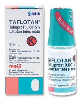
TAFLUTAN


Tafluprost 0.0015% is a prostaglandin analogue which was co-developed by Santen with Asahi Glass Co., Ltd (Tokyo, Japan) for the treatment of glaucoma and elevated intraocular pressure (IOP). Unit-dose, preservative- free eyedrops and in combination with timolol are now also available.  

The recommended dose is one drop of tafluprost in the conjunctival sac of the affected eye(s) once daily in the evening.


PHARMACOLOGY:

Mechanism of action=

Tafluprost acid is a fluorinated prostaglandin F2α (PGF2α) analogue. Tafluprost is a prodrug of the active substance, tafluprost acid, a structural and functional analogue of PGF2α. Tafluprost acid is a selective agonist at the prostaglandin F-receptor, increasing outflow of aqueous humor via the uveoscleral pathway and thus lowering IOP.

Other PGF2α analogues with the same mechanism of action include latanoprost and travoprost.


Pharmacokinetics=

Tafluprost is a prodrug ester prostaglandin F2α-analog designed to expedite the corneal penetration of the drug, which is then hydrolyzed by corneal esterases to produce the carboxylic acid active metabolite. The product, tafluprost acid, can then be taken up by the aqueous humor to therapeutically relevant levels.

Onset of action is 2 to 4 hours after application, the maximal effect is reached after 12 hours, and ocular pressure remains lowered for at least 24 hours.

Tafluprost acid is inactivated by beta oxidation to 1,2-dinortafluprost acid, 1,2,3,4-tetranortafluprost acid, and its lactone, which are subsequently glucuronidated or hydroxylated. The cytochrome P450 liver enzymes play no role in the metabolism.

ADVERSE EFFECTS:

The most common side effect of tafluprost is conjunctival hyperemia, which occurs in 4 to 20% of patients. Less common side effects include stinging of the eyes, headache, and respiratory infections. Rare side effects are dyspnea (breathing difficulties), worsening of asthma, and macular oedema.

Tafluprost causes changes to pigmented tissues, leading to increased pigmentation of the iris, periorbital tissue (eyelid) and eyelashes. Before treatment is initiated, patients should be informed of the possibility of eyelash growth, darkening of the eyelid skin and increased iris pigmentation. Some of these changes may be permanent, and may lead to differences in appearance between the eyes when only one eye is treated.

Usually, the eyelash and pigmentary changes resolve after discontinuation of the drug.

To reduce the risk of darkening of the eyelid skin patients should blot off any excess solution from the skin. Nasolacrimal outflow occlusion or gently closing the eyelid after administration may reduce the systemic absorption of products administered via the ocular route.

Contact lenses should be removed prior to the administration of tafluprost, and may be reinserted 15 minutes following administration.

Caution is recommended in patients with known risk factors for iritis/uveitis and should generally not be used in patients with active intraocular inflammation.

Macular edema, including cystoid macular edema, has been reported during treatment with prostaglandin F2α analogues. These side-effects usually occur in aphakic patients, pseudophakic patients with a torn posterior lens capsule or anterior chamber lenses, or in patients with known risk factors for macular edema. Therefore, caution is recommended when using tafluprost in these patients.

STUDIES:

In a review performed by Keating, tafluprost was at least as effective as latanoprost ophthalmic solution 0.005 % in Asian patients with primary open-angle glaucoma or ocular hypertension. The efficacy of tafluprost ophthalmic solution 0.0015 % was maintained in the longer term. [1]

A study by the Tafluprost Multi-center Study Group and others, found the agent to be effective in lowering IOP in normal-tension glaucoma (NTG) patients. Mean IOP changes from baseline were -4.0 +/- 1.7 mmHg in tafluprost administered patients and -1.4 +/- 1.8 mmHg in Placebo administered patients at 4 weeks, with a statistically significant difference (p<0.001). [2]

A study by Nakano et al., to evaluate the efficacy and safety of tafluprost in NTG with IOP of 16 mmHg or less, found the IOP in the study eyes versus fellow eyes were 10.2 ± 1.6 versus 12.1 ± 1.5 mmHg at week 12 of treatment. The IOP difference between the study eyes and the fellow eyes was statistically significant (P < 0.0001, Student's t test). [3]

Hoy has reported good IOP control with the tafluprost/timolol combination (Taptiqom). [4]


REFERENCES:

  1. Keating GM. Tafluprost Ophthalmic Solution 0.0015 %: A Review in Glaucoma and Ocular Hypertension. Clin Drug Investig. 2016 Jun;36(6):499-508. doi: 10.1007/s40261-016-0413-z. PMID: 27225879.
  2. Kuwayama Y, Komemushi S; Tafluprost Multi-center Study Group. [Intraocular pressure lowering effect of 0.0015% tafluprost as compared to placebo in patients with normal tension glaucoma: randomized, double-blind, multicenter, phase III study]. Nippon Ganka Gakkai Zasshi. 2010 May;114(5):436-43. Japanese. PMID: 20545217.
  3. Nakano T, Yoshikawa K, Kimura T, Suzumura H, Nanno M, Noro T. Efficacy and safety of tafluprost in normal-tension glaucoma with intraocular pressure of 16 mmHg or less. Jpn J Ophthalmol. 2011 Nov;55(6):605-13. doi: 10.1007/s10384-011-0082-7. Epub 2011 Aug 27. PMID: 21874307.
  4. Hoy SM. Tafluprost/Timolol: A Review in Open-Angle Glaucoma or Ocular Hypertension. Drugs. 2015 Oct;75(15):1807-13. doi: 10.1007/s40265-015-0476-9. PMID: 26431840.


GLAUCOMA FIELD DEFECT CLASSIFIER (GFDC)

  Glaucoma Field Defect Classifier  ( GFDC ) is a web application that automates glaucoma grading based on Hodapp-Parish-Anderson (HPA) cri...