Monday, July 29, 2019

COMING SOON

THE FLAMMER SERIES 
ON THE GLOG

A SERIES OF POSTS DEDICATED TO 
PROFESSOR JOSEF FLAMMER


PROGRAM:
1st August 2019
Prof Josef Flammer: Biographical sketch of a medical innovator.
Author= Dr Ronald Gerste, Germany.
1st August 2019
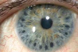
Prof Flammer, a visual experience.
8th August 2019
The Flammer Syndrome.
Author= Saleha Hasan, India.
15th August 2019
Ocular Blood Flow in glaucoma.
Author= Ghuncha Khatoon, India.
22nd August 2019
Topic not yet decided.
Author= Dr S Shoeb Ahmad.
29th August 2019
Normal tension glaucoma.
Author= Dr Ronald Gerste, Germany.

Sunday, July 28, 2019

ARGON LASER PERIPHERAL IRIDOPLASTY


Guest author
SEHRISH
Ajmal Khan Tibbiya College
Aligarh, India


INTRODUCTION

Iridoplasty, also known as gonioplasty, uses low-energy laser burns to the peripheral iris in order to widen the anterior chamber angle and/or break peripheral anterior synechiae. Patients requiring laser iridoplasty are most often diagnosed with plateau iris syndrome, either by ultrasound biomicroscopy or follow up gonioscopy that demonstrates a narrow angle after laser peripheral iridotomy.

During iridoplasty, the laser light is converted to heat that causes contraction of stromal collagen, which is primarily responsible for the immediate anatomical change. Later alterations include a proliferation of fibroblasts with the formation of a contraction membrane. Careful technique (see Step-by-Step Technique for Iridoplasty) is important, because overtreatment can lead to coagulative necrosis of the blood vessels.

HISTORY

Krasnov and Kimbrough’s attempts to modify the peripheral iris had some success; however,the outcomes were limited by technique and instrumentation.

Kimbrough et al described a technique for direct treatment of 360° of the peripheral iris through a gonioscopy lens, and termed the procedure gonioplasty. 

The current use of argon lasers has led to a refinement in technique that has increased both anatomical and clinical success.

ARGON LASER PERIPHERAL IRIDOPLASTY (ALPI)

Argon laser peripheral iridoplasty is a useful procedure to eliminate appositional angle closure resulting from mechanisms other than pupillary block. For those eyes with angle closure originating at an anatomic level posterior to iris, such as- plateau Iris, lens-induced angle closure, malignant glaucoma, central retinal vein occlusion etc.

Argon laser peripheral iridoplasty is often useful in these cases to further open the angle. It can be used to break an acute attack of angle closure glaucoma and relieve appositional angle closure secondary to plateau iris syndrome, or lens-related angle closure, and to widen the angle prior to argon laser trabeculoplasty treated as necessary if a postlaser IOP rise occurs.

CHARACTERISTICS OF ARGON

Phocoagulative ( lower energy & longer exposure)

Iris color (Pigment density) is the most important factor.

   (a) Light brown colour: 600-1000mW with a spot size of 50ûm and a shutter speed of 0.02-0.05 second
   (b)  Dark brown colour: 400-1000mW, spot size of 50ûm and a shutter speed 0.01 second
    (c) Blue Iris colour: 200-400mW, spot size 200ûm, speed 0.1 second

INDICATIONS


  1. Acute angle closure glaucoma
  2. Chronic angle syndrome
  3. Plateau iris syndrome
  4. Angle closure due to size or position of lens
  5. Iris bombe
  6. Adjunct to laser trabeculoplasty
  7. Malignant glaucoma
  8. Fellow eye
  9. Nanophthalmos
  10. Aphakic or pseudophakic pupillary block 
  11. Incomplete surgical iridectomy
  12. Subluxated Crystalline lens
  13. Aqueous misdirection syndrome
  14. Pigmentary glaucoma
  15. ACIOL implant 

CONTRAINDICATIONS


  1. Severe and extensive corneal edema or opacity
  2. Flat anterior chamber
  3. Synechial angle closure


TECHNIQUE

STEP-BY-STEP TECHNIQUE FOR IRIDOPLASTY

STEP 1: The informed consent for iridoplasty includes an explanation of potential side effects such as:
Pain/discomfort
• Inflammation
• Elevated IOP
• Changed pupillary shape/size
• Possible need for retreatment

STEP 2 :Pretreat the patient with:
One drop of pilocarpine 2%
• One drop of brimonidine or apraclonidine
• One drop of proparacaine

STEP 3: Set up laser:
Power—300 to 500 mW (higher if needed)
• Spot size—300 to 500 µm
• Duration—300 to 500 milliseconds

STEP 4
Place Genteal gel (Novartis Ophthalmics, Inc., Duluth,GA), Refresh Celluvisc (Allergan, Inc., Irvine, CA), or another clear lubricant on the single-mirror lens (or the Abraham lens, if you choose) and position the lens over the eye.

STEP 5
Treat the peripheral iris without encroaching on the trabecular meshwork.
• Increase the laser power as needed to cause the tissue to contract without forming bubbles or releasing pigment.
• Treat 360º.

STEP 6
Remove the lens and clean off the eye.
• Instill one drop of prednisolone acetate 1%.
• Recheck the IOP in 1 hour.
• Send the patient home with instructions to administer one drop of Prednisolone q.i.d. for 4 days
• Follow up in 1 week.
                                       
After the procedure, the eye receives a drop of a topical steroid or NSAID. The surgeon checks the IOP 1 hour after treatment. At the 1-week follow-up visit, check the patient’s IOP and perform gonioscopy to re-examine the anterior chamber angle. Retreatment may be indicated in some cases and will consist of either overlapping the spots or adding a row of applications to the initial treatment.

POST-OPERATIVE TREATMENT

Immediately after the procedure, the patient is given a drop of topical steroid and apraclonidine or brimonidine. Gonioscopy should be performed to assess the effect of the procedure immediately if pilocarpine has not been used. If it has, it is better to evaluate the success of the procedure at a sub-sequent visit. Patients are treated with topical steroids 4--6 times daily for 3 to 5 days. Intraocular pressure is monitored postoperatively as after another anterior segment laser procedure and patients treated as necessary if a post-laser IOP rise occurs.

COMPLICATIONS

Mild postoperative iritis
Iris atrophy
Ocular irritation.
Iridoplasty is often performed on patients with extremely shallow peripheral anterior chambers, diffuse corneal endothelial burns may occur.
During laser iridotomy, endothelial burns seen during ALPI are larger and much less opaque.
Endothelial burns present a problem early in the procedure, they may be minimized by placing an initial contraction burn more centrally before placing the peripheral burn (kriss-kross iridoplasty).
          A transient rise in IOP can occur as with other anterior segment laser procedures.    
          Lenticular opacification has not been reported.
        When IOP is rapidly reduced in acute primary angle closure by ALPI, decompression     retinopathy can rarely occur. 
         Urrets-Zavalia syndrome.
         Recurrence of angle-closure.

NEED FOR RETREATMENT

Although ALPI is highly successful long-term in eyes with plateau iris, patients need to be followed closely for recurrence of appositional closure, and if this develops, may require retreatment.

Patients should be observed gonioscopically at regular intervals and further treatment given if necessary. 

This is most common in a patient in whom the mechanism of the glaucoma is lens-related or as the lens enlarges over time.

Retreatment is only occasionally needed in patients with plateau iris, whereas those with intumescent lenses usually undergo cataract extraction.

CONCLUSION
  
Argon laser iridoplasty is a safe and effective procedure for patients with narrow angles and/or plateau iris syndrome whose angles remain narrow after laser iridotomy.

When properly performed, the procedure consistently delivers a long-term benefit to individuals with plateau iris syndrome. Patients with acute ACG may also profit from iridoplasty in cases where immediate peripheral iridoplasty/dotomy cannot be performed. 

A precise technique and an attention to detail are keys to successful iridoplasty, and ophthalmologists should be familiar with the finer points of performing this laser procedure. 


Thursday, July 25, 2019

CENTRAL CORNEAL THICKNESS AND GLAUCOMA


Guest author

ALIYA

Ajmal Khan Tibbiya College
Aligarh
India 


INTRODUCTION

  • According to the 11th century Arab scholar, Abu Al-Hassan Tabarri, a firm or hard eye was an indication of glaucoma. Over the years intraocular pressure (IOP) has become the bedrock in the positive and negative association with glaucoma. On one hand IOP is regarded as a surrogate factor in the assessment of glaucoma, while on the other, certain conditions such as ocular hypertension and normal-tension glaucoma question the importance of this marker to diagnose glaucoma.
  • There are a number of instruments developed over the years to measure IOP. However, despite all our advances, Goldmann Applanation Tonometry (GAT) continues to be labeled as the “gold standard” to assess this parameter. Unfortunately, GAT is fraught with a number of errors such as improper technique; patient factors such as cooperation during measurement and tight neckties; size of the semi-circles (Wider menisci cause falsely higher pressure estimates. Improper vertical alignment will also lead to a falsely high IOP estimate.); corneal factors such as corneal thickness, curvature and astigmatism; calibration errors and error due to prolonged contact.
  • GAT is dependent on a standard central corneal thickness (CCT), leading to errors in IOP measurement if the CCT is beyond the standard values (explained later). Unfortunately, the mean CCT in healthy human eyes varies by ethnoracial characteristics. This produces controversial confounding factors in the measurement of IOP by GAT.
  • Apart from being a factor in IOP measurement, CCT is also regarded as a risk factor for development of glaucoma and also associated with glaucoma severity and progression.
  • Most clinical practice guidelines do recommend evaluation of CCT in the initial assessment of all glaucoma patients. This post takes a look at CCT and how it affects glaucoma diagnosis and management.

CCT AND OCULAR HYPERTENSION:


  • The Ocular Hypertension Treatment Study (OHTS) has shown that thin CCT is the most important predictive factor for the development of primary open angle glaucoma (POAG).
  • However, there are indications that the significance of CCT as a risk factor for glaucoma could have been overestimated in OHTS.
  • OHTS assessed patients who had high IOP at baseline (without other glaucoma characteristics), this led to a selection bias towards individuals who had thicker CCT.
  • 25% patients had CCT above 600µ, so a significant number of participants in OHTS had corrected IOP of less than 21 mmHg.
  • Conversely, individuals with thin CCT would have their IOP overestimated during eligibility screening.
  • These factors would lead to overestimation of the significance of CCT as a risk factor for the development of glaucoma.
  • Although these limitations are important, CCT has been established as an important factor to consider in the risk stratification of OHT patients.
  • The OHTS prediction model was independently validated in the European Glaucoma Prevention Study (EGPS) and also in the pooled OHTS-EPGS data.
  • Every 40µ decrease in CCT was associated with a two-fold increase in the risk of developing glaucoma over 5 years.
  • CCT measurement would minimize the overtreatment of OHT patients with thick corneas.
  • In patients with thin CCT and borderline IOP, initiating treatment can be considered keeping in view other relevant risk factors.
  • It needs to be emphasized that IOP alone (even when adjusted for CCT) is not a reliable screening tool for POAG.

CCT AND GLAUCOMA


  • The role of CCT in the clinical management of patients with established glaucoma is less well recognized (compared to OHT).
  • Prevalence of POAG was significantly higher in individuals with thin (compared to normal or thick) CCTs in the Los Angeles Latino Eye Study.
  • Studies in West Africa, East Asia and China did not show any significant difference in CCT in glaucomatous versus non-glaucomatous eyes.
  • Studies have shown eyes with NTG are associated with thinner CCT than eyes with POAG and normal eyes.
  • Pseudoexfoliative glaucoma and PACG have been found to be associated with thin corneas in some studies but refuted in others.
  • It is controversial whether any significant association exists between CCT and glaucoma progression or glaucoma severity.
  • Early Mainfest Glaucoma Trial (EMGT) did show thinner CCT to be associated with glaucoma progression.
  • However, other landmark glaucoma trials such as Advanced Glaucoma Intervention Study (AGIS), Collaborative Initial Glaucoma Treatment Study (CIGTS) and Collaborative Normal Tension Glaucoma Study (CNTGS) did not analyze the impact of CCT on glaucoma.
  • A few other recent studies have not found any significant association between CCT and glaucoma.

POSSIBLE MECHANISMS FOR THE ASSOCIATION BETWEEN CCT AND GLAUCOMA


(1) Tonometry artifact:

  • GAT is based on an average CCT of 500µ.
  • Ehlers cannulated the anterior chamber to obtain true IOP levels and found the most accurate GAT results were obtained with CCT of 520µ.
  • Every 100µ of deviation in CCT would produce an error of 7 mmHg in IOP measurement.
  • A thick cornea causes an artefactually increase IOP reading, while a thin cornea produces an artefactually reduced IOP reading.
  • Corneal compensated IOP is either independent or weakly dependent on CCT.
  • Certain tonometers such as Ocular Response Analyzer (ORA) and Corvis tonometer are able to compensate for CCT.
  • Other tonometers such as Dynamic Contour Tonometry and Transpalpebral Tonometry have their own drawbacks.
  • Linear correction factors for IOP based on CCT have been proposed, however based on mathematical models, the relationship between CCT and IOP is non-linear and complex.
  • So far no adequately validated correction algorithm for GAT measurements has been provided.

(2) Biological risk factor:

  • While certain studies have shown increased movement of lamina cribrosa in response to IOP changes in individuals with thin CCTs, others have refuted this.
  • Another hypothesis suggests that a thin cornea increases the exposure of the trabecular meshwork to oxidative damage (a study reported an inverse correlation between CCT and partial pressure of oxygen in the anterior chamber).

(3) Genetic associations:

  • CCT is often inherited, with heritability estimates for CCT ranging from 88-95%.
  • Apparently a number of genes are responsible for inheritance of CCT.
  • Again, while there are studies suggesting the role of mutations and single nucleotide polymorphisms, others did not find any genetic correlation between CCT and POAG.

GLAUCOMA TREATMENT


  • Ocular hypotensive medications have been shown to alter corneal thickness.
  • Most studies have reported an association between the use of topical prostaglandin analogues (PGA), including latanoprost and tafluprost, and a significant decrease in CCT, possibly by modifying the extracellular matrix.
  • However, there are other studies which did not find any correlations between PGA use and CCT.
  • Topical beta-blockers have also been reported to cause reversible increase in CCT.
  • Topical carbonic anhydrase inhibitors can cause irreversible corneal decompensation and increased corneal thickness in eyes with underlying corneal disease. However, this effect is not seen in healthy corneas.
  • Conversely, there appears to be an effect of corneal thickness on the response of glaucoma treatment.
  • The OHTS reported that thicker CCT was associated with significantly less decrease in IOP in response to anti-glaucoma treatment.
  • In a study of Selective Laser Trabeculoplasty for POAG and OHT, thinner corneas were associated with comparatively higher IOP reduction.

VARIABILITY IN CCT

  • CCT measurements vary with race, sex, age and other environmental factors, which may confound the relationship between glaucoma and corneal thickness.
  • African (518-533µ), Indian (511-514µ), Mongolian (495-514µ) and Japanese (517-532µ) individuals were found to have thinner corneas compared to white (542-558µ), Hispanic (547µ), Korean (554µ) and Chinese (540-542µ) persons in population-based studies.
  • Racial differences in CCT would need to be considered in the interpretation of IOP and may explain the higher rates of glaucoma in certain populations.
  • Wang et al. found that the variation in CCT accounted for upto 29.4% of the increased risk of glaucoma seen among African-American persons in a large multiethnic population in Northern California.
  • In the OHTS, African-American ethnicity was predictive of POAG in univariate analysis, though it was not significant in the multivariate analysis after inclusion of CCT in the model, suggesting the increased susceptibility of this group of individuals to glaucoma can be partially explained by their thin CCT.
  • Most epidemiological studies found men to have thicker corneas.
  • CCT varies with age, with a small but significant inverse relationship between CCT and age (range: 2-10microns per decade). This is comparable to longitudinal data from OHTS (6microns per decade).
  • It can be surmised that it would take 20 years for the change in CCT to be clinically significant and to warrant remeasuring.
  • CCT is also influenced by environmental factors. Dry eyes and long term contact lens wear are associated with a decrease in CCT, whereas occupation indoors and sleep are associated with an increase in CCT.
  • The use of various devices which use different modalities to measure CCT has also resulted in variable results of CCT.
  • Most landmark glaucoma studies such as OHTS, EMGTS and EGPS used ultrasound pachymetry.
  • There are other instruments also being used to measure CCT. These include: Scheimpflug imaging, optical coherence tomography, specular microscopy and scanning slit topography.
  • It is ideal to use the same device for the same patient for every CCT measurement.

CORNEAL BIOMECHANICS

  • Corneal biomechanics may have a more significant impact on IOP measurement errors than CCT which is merely a corneal dimension and not a biomechanical property.
  • CCT is regarded as a correlate of corneal rigidity, but this is only applicable to structurally normal Cornea.
  • An edematous Cornea has lower corneal rigidity and therefore, despite increased thickness will have lower GAT readings.
  • Conversely, a scarred cornea, although thinner, has increased rigidity and will demonstrate artefactually higher IOP readings.
  • Eyes undergoing corneal cross linking show higher IOP on an average 12 months after the procedure, apparently due to higher corneal rigidity.
  • Measurements of Corneal biomechanical properties, such as corneal hysteresis, corneal resistance factor and corneal constant factor can be obtained by the Ocular Response Analyzer and the Corvis ST tonometer.
  • These parameters may be better correlated with overall globe biomechanics than CCT and may help to explain the susceptibility of some optic nerve heads to damage as a result of IOP variations.


GLAUCOMA FOLLOWING INFANTILE CATARACT SURGERY

  Cataract surgery, especially infantile cataract surgery, is associated with a higher incidence of glaucoma post-operatively. Choe et al ...