Wednesday, June 22, 2016

AQUEOUS FORMATION

Glaucoma is a neurodegenerative disorder attributed to a multifactorial etiology. 
The major factor responsible is supposedly a defect in the balance between aqueous humor (AH) production and outflow through the eye, thus affecting intra-ocular pressure (IOP). This abnormal IOP is the main risk factor for optic nerve damage in glaucoma patients.
Thus, understanding of glaucomatous optic nerve degeneration (GOND) requires a clear knowledge of AH physiology and IOP.  
Currently, IOP is assumed to be a major risk factor and perhaps the only factor which can be controlled
IOP can be controlled medically, by laser, surgery or other means. Among the pharmacologic agents, most decrease aqueous humor (AH) production or increase the aqueous outflow through the unconventional uveoscleral route. Among these agents, an important group is one of carbonic anhydrase inhibitors (CAIs). This group includes both systemic (oral and intravenous) as well as topical agents. CAIs reduce aqueous formation and play a role in improving ocular blood flow.
Thus, CAIs form an important group of agents which need to be studied in more detail to understand their indications, mode of action, side-effects and any new developments which can be useful for the practicing clinician.
AQUEOUS FORMATION AND ROLE OF CARBONIC ANHYDRASE:
There are a number of theories to explain the formation of AH. These include: the dialysis theory; active transport/secretion theory and the ultrafiltration theory. According to the active transport theory, AH formation is an energy-dependent process. This energy-dependent process is able to move substances across a concentration gradient in a direction opposite to what would be expected by passive mechanisms alone.
The presence of higher concentrations of ascorbate, lactate and some amino acids in AH, compared to plasma levels, suggest the role of active transport. Studies have shown that the ciliary epithelium pumps substances against their concentration gradient, so that their levels are higher in AH compare to that in plasma. This is achieved by the utilization of certain enzyme systems in the ciliary epithelium.
Sodium-Potassium activated adenosine triphosphate (Na-K ATPase ) pumps Na+ across the cell membrane. While Cl- passively follows it to maintain electrical neutrality. Another enzyme is Carbonic Anhydrase (CA). This is found in the cell membrane and cytoplasm of both non-pigmented and pigmented epithelia of the ciliary body. The enzyme catalyzes the following reaction=
CO2 + OH-  -- H+ + HCO3-
Thus, the levels of bicarbonate are found to be higher in AH compare to plasma (34 mmol/kg H2O vs. 24 mmol/kg H2O). CA plays an indirect role in AH formation by providing hydrogen or bicarbonate ions for an intracellular buffering system.
Humans have 16 isoforms of CA (with 13 having catalytic activity). Human eyes contain CA I, II and IV. CA I and II are present in the cytosol while CA IV is membrane bound. CA IV was detected in the ciliary processes of mice, but not in the human ciliary processes. It was also found that patients who lacked CA II had no effect of intravenous acetazolamaide on IOP. Thus, it is likely that CA II plays a major role in AH secretion.
CA is an ubiquitous enzyme found widespread in nature among animals and photosynthesizing organisms including bactieria. It has also been found recently in some non-photosynthesising bacteria. There are 5 genetically distinct families of CAs. These include: CA α, β, γ, δ, ζ. All of them are metalloenzymes. CA α, β, δ use Zn++ ions at the active site; CA γ has Fe++ ions (but is also active with Zn++ and Co++), while CA ζ uses Cd++ or Zn++.
CA α is usually present as monomers, but can rarely occur as dimers. CA β can occur as dimers, teramers or octamers; CA γ are trimers; whereas δ and ζ are probably monomers.
CAs also play a role in respiration and transport of CO2/bicarbonate, pH and CO2 homeostasis, electrolyte secretion, biosynthetic reactions (gluconeogenesis, lipogenesis and ureagenesis), tumorigenecity and photosynthesis. They may also be responsible for other biosynthetic reactions in some bacteria, algae and plants as well as, in CO2 fixation in diatoms. 
AHMAD'S MODIFIED TRABECULECTOMY TECHNIQUE


Since its introduction in the 1960s, trabeculectomy has remained the gold standard of glaucoma filtering surgeries (GFS). However, this procedure remains unpredictable with results varying from patient to patient and even from case to case done by the same surgeon. It is also known that the more the risk factors for the procedure, the higher the cumulative effect on the outcome. Other weaknesses of this procedure include the safety profile, complications such as short- and long-term risks of infection (endophthalmitis, blebitis), ocular hypotony and others. Patients with cystic blebs carry a lifetime risk of bleb related infections.
The key factor which determines the success of trabeculectomy is the characteristic of the filtering bleb. Since, bleb related complications usually influence the outcome of glaucoma filtering surgeries we have developed some modifications to the classical trabeculectomy procedure. Our method, is aimed primarily at overcoming the problems of bleb leakage and poor quality of blebs. This technique targets to achieve the “much desired diffuse, non-cystic bleb” through promoting posterior flow by making an innovative “spout” configuration in the sclerostomy, tight suturing of the scleral flap anteriorly and better healing by leaving a cuff of conjunctiva near the limbus.

SURGICAL TECHNIQUE:
A conjunctival flap is fashioned. Unlike the usual fornix-based flaps which are flush with the cornea, we start the peritomy about 0.5mm posterior to the limbus leaving a margin of conjunctiva there.
We routinely use Mitomycin-C (Kyowa Hakko Kirin Co., Japan). The agent is diluted to a concentration of 0.02% solution. Subsequently, gel sponges soaked in the solution are applied to the episcleral tissues. The sponges are kept for 2-3 minutes, with the conjunctival flap blanketed over them by holding the edges away from the sponges. After 2-3 minutes, the sponges are removed and the area irrigated with nearly 20ml of balanced salt solution (BSS).
A bevelled limbal stab incision is made at the 9 o’clock region after the Mitomycin-C is washed out. Using a 15o knife, a square scleral flap, measuring 4mm x 4mm is fashioned, hinged at the limbus. The dissection is completed using a crescent knife. Subsequently, a stab incision is made into the anterior chamber just behind the hinge of the scleral flap. Now, a Kelly-Descemet punch (1 mm diameter) is used to cut the deep sclero-corneal layer in order to perform the sclerostomy. Usually 2-3 overlapping cuts are made. Subsequently, we go back to the centre of the sclerostomy and, tilting the punch downwards, make a "half-thickness" cut in the sclera at the posterior lip of the sclerostomy. This produces a spout-like configuration in the center allowing aqueous humor to flow posteriorly, rather than towards the sides.
Block arrows show sclerostomy and open arrow reveals pigment coming out through the "spout".


Dimensions of the trabeculectomy


A peripheral iridectomy is then made. The scleral flap is sutured with 5 10/0 nylon sutures. We keep the anterior 2 sutures on either side relatively tight while the remaining 3 are kept loose. BSS is injected from the limbal stab incision to titrate the flow. The sutures are adjusted to have a smooth flow through the fistula without collapse of the anterior chamber on applying pressure. This modification has two advantages. One, it directs flow more posteriorly and second, in the event the patient requires laser suture lysis, it is easier to lyse the anterior 2 tight sutures.
Final appearance of the bleb. Open arrows showing the position of the peritomy.


CONCLUSION:
The advantages of this procedure are: (i) Aqueous is directed posteriorly by the 2 tight anterior sutures and the scleral-spout. This prevents bleb leakage and formation of cystic blebs.  (ii) There is good healing of the conjunctival edges. This prevents bleb leakage also.
(Kindly google for: Ahmad's Modified Trabeculectomy Technique )

Monday, June 20, 2016


IMAGE LIBRARY


KK1M, Queen Elizabeth Hospital, 8-10 September 2017

Image of Krukenberg Spindle (c) Dr Daljit Singh


 Mouse RGCs: Nikon Macrophotography Contest 2016

Phacoemulsification Camp at Queen Elizabeth Hospital, KK. 16-18 September 2016


Secondary Eye Care Course, Queen Elizabeth Hospital, 29th August 2016
Pfizer Glaucoma Talk at Queen Elizabeth Hospital, 11th October 2016
 

Allergan launches Lumigan 0.01% in Malaysia. 22.7.2016

Borneo Glaucoma Workshop, Miri, Sarawak, April, 2016
Dr Francisco Goni, Allergan Talk, Kota Kinabalu, 4th April, 2016
Asia pacific Glaucoma Congress, Chiang Mai, Thailand. 14-16.7.2016
Dr Michael Coote, UKM-SSYQOL Symposium, Kuala Lumpur, October 2015
6th World Glaucoma Congress, Hong Kong
Dr Robert Fechtner, Alcon Glaucoma Talk, Kuala Lumpur, March 2016

Glaucoma Walk, Kota Kinabalu, 2014
Glaucoma Walk, Kota Kinabalu, 2016


Monday, June 13, 2016

AN ODE TO NVG
(A poem !)
--------------------------------------------------------------------------------------------
Once I met a patient who had proliferative diabetic retinopathy and neo-vascular glaucoma. 
Touched by her suffering I wrote this poem......

Postscript: An ODE to NVG

She was a girl just turned twenty-one;
A future nurse, when her course was done.
Her father has a history of DM;
But it does not cause him any problem.
The killer genes have hit her hard;
Attacking her eyes, which have no guard.
New vessels growing all over the disk;
There is also NVE, to increase the risk......
Read the complete poem at the following link >>>>>>>


Thursday, June 9, 2016

PIGMENTARY GLAUCOMA
 
Pigment dispersion in the eye could be part of the natural process of aging.
The infant eye, especially the trabecular meshwork, is non‑pigmented. However, with aging, various grades of pigmentation might become visible.
In most cases, pigment dispersion is innocuous and does not cause any pathological changes in the eye.
This condition is known as Primary Pigment Dispersion Syndrome (PPDS). It is characterized by pigment deposition throughout the anterior segment.
The common sites of pigment deposition are: the corneal endothelium, iris surface, lens zonules and capsules, as well as the anterior chamber angle.

PPDS can convert to open angle glaucoma over time. Thus, it is important to monitor such cases carefully.
-------------------------------------------------------------------------------------------------------------------
CAUSES OF TRABECULAR PIGMENTATION
1. P seudoexfoliation & pigment dispersion
2. I ritis
3. G laucoma (following acute angle closure)
4. M elanosis of angles (oculodermal melanosis)
5. E ndocrine (diabetes & Addison's disease)
6. N evus (Cogan-Reese syndrome)
7. T rauma
---------------------------------------------------------------------------------------------------------------------
We recently reported about a patient who also had peripheral retinal pigmentary abnormalities. Please check link:

http://www.ncbi.nlm.nih.gov/pmc/articles/PMC4860978/


Wednesday, June 8, 2016

THE BIOCHEMICAL MECHANISMS OF GLAUCOMATOUS NEURODEGENERATION

There is ample evidence from studies done at various laboratories world-wide, as well as, from clinical trials, that biochemical mechanisms play an important role in the causation of glaucomatous optic nerve degeneration (GOND).

These biochemical mechanisms include excitatory aminoacids, caspases, protein kinases, oxygen free radicals, nitric oxide, TNF alpha, neurotrophins and metalloproteins.
This is an article which covers the salient points of these mechanisms in brief.

http://www.ncbi.nlm.nih.gov/pmc/articles/PMC4741173/

GLAUCOMA DEMOGRAPHICS
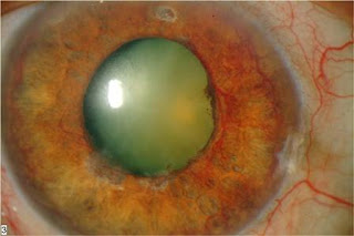
  •  Etymology: The word "glaucoma" comes from the Greek γλαύκωμα, a derivative of γλαυκóς, which commonly described the color of eyes which were not dark (i.e. blue, green, light gray). Eyes described as γλαυκóς due to disease might have had a gray cataract in the Hippocratic era, or, in the early Common Era, the greenish pupillary hue sometimes seen in angle-closure glaucoma.
  • Glaucoma is the second leading cause of blindness in the world, according to the World Health Organization.
  • Estimates put the total number of suspected cases of glaucoma at over 60 million worldwide.
  • It is estimated that over 3 million Americans have glaucoma but only half of those know they have it. 
  • In the U.S., more than 120,000 are blind from glaucoma, accounting for 9% to 12% of all cases of blindness.
  • Blindness from glaucoma is 6 to 8 times more common in African Americans than Caucasians.
  • Glaucoma accounts for over 10 million visits to physicians each year.
  • http://www.glaucoma.org/glaucoma/glaucoma-facts-and-stats.php
  •  
  • Because of the aging of the world population, one estimate shows that the number of persons with glaucoma will increase to79.6 million in 2020  
  • Our analysis estimates that there will be a substantial increase in the number of persons with POAG in the United States, from 2.71 million in 2011 to 7.32 million in 2050.
  • http://iovs.arvojournals.org/article.aspx?articleid=2126921
  •  
  • Overall, the results demonstrate that glaucoma is responsible for approximately 5.2 million blind (15% of the total burden of world blindness).
  •  http://www.who.int/blindness/publications/glaucoma/en/ 

A 40-Year Forecast for POAG

In 2011, 2.71 million people in the United States had POAG, with the highest estimated number among populations aged 70 to 79 years (31%), women (53%), and non-Hispanic whites (44%). The largest demographic group is non-Hispanic white women.
In 2050, an estimated 7.32 million people in the United States will have POAG, with the highest number among populations aged 70 to 79 years (32%), women (50%), and Hispanics (50%). The largest demographic group is Hispanic men.

Despite the high prevalence of POAG in African Americans and Hispanics, the largest group in the United States with POAG is among older non-Hispanic white women (2011), but is expected to shift to Hispanic men over the next few decades.
http://www.aao.org/eye-disease-statistics
  • The Eye Disease Prevalence Research Group developed estimates of glaucoma prevalence from data that included multiple US-based and international studies. The group estimated that open-angle glaucoma prevalence increased from 0.7% at age 40 to 7.7% for those over age 80.3 Self-reported epidemiologic data from the National Health Interview Survey found glaucoma prevalence increasing from 0.2% at age 18 to over 10.7% for those over 75.3 The growth in the oldest-old is important, as the prevalence of glaucoma triples in those over 75 compared to those aged 65.
  • https://www.reviewofoptometry.com/article/the-changing-and-challenging-epidemiology-of-glaucoma

  •  


PHARMACOLOGIC TRABECULECTOMY 
“Pharmacologic or medical trabeculectomy” is a term first used in the 1970s. This describes a process by which the trabecular meshwork can be biochemically manipulated in order to reduce the outflow resistance and, thus, the intraocular pressure (IOP). 

In cases of primary open angle glaucoma and normal-tension glaucoma , the IOP can be controlled by either reducing the inflow of aqueous humor into the eye or by increasing the outflow. Most of the medications in use now reduce aqueous production or the aqueous egress through the unconventional uveoscleral pathway. 

According to one school of thought, decreasing the aqueous production and thus reducing aqueous outflow through the trabecular meshwork will progressively be detrimental to the trabecular function. This could be further accelerated by concurrent usage of prostaglandin analogs, which direct aqueous away from the meshwork to the ciliary body. 

In order to overcome this, and to develop a new line of approach to the management of glaucoma, interest is focusing on the trabecular outflow pathways, and the modulation of which, can be used to reduce IOP. 

In this review we take a look at the agents that have been investigated to improve the facility of aqueous outflow.
 
AQUEOUS HUMOR OUTFLOW DYNAMICS

Aqueous humor outflow occurs through the conventional and unconventional pathways. 
With aging, the latter becomes less active so that the conventional pathway remains the primary mechanism of aqueous humor outflow. 
An abnormality of this pathway contributes significantly to disordered aqueous humor dynamics and consequent rise in intraocular pressure seen in primary open angle glaucoma and ocular hypertension. Recently, the ocular lymphatics have been implicated in aqueous humor outflow. 
The trabecular meshwork is now understood to be a complex organization of structures, which are controlled by various biomechanical and biochemical mechanisms. Among others, these include the actinomyosin cytoskeletal system, extracellular matrix, intracellular signaling responses mediated by protein kinase C, Rho/Rho kinase, and other biologic factors. 
The following review describes the various pathophysiologic mechanisms involved in aqueous humor dynamics.

Sunday, June 5, 2016

Glaucoma Specialty Club: A start


This glaucoma blog (or "glog", as I shall henceforth call it) has been made for those ophthalmologists who have interest in glaucoma. 

We can discuss about stuff which appeals us. Be it basic sciences, diagnostic methods or management strategies

This brings us to the question of who I am? I am an ophthalmologist with almost 20 years of experience working in many hospitals across Asia. My interest in glaucoma has led me to re-visit old theories and explore new areas of research. What specially stimulates me are the "biochemical mechanisms of glaucoma causation" and "newer methods to treat glaucoma".

On this glog I shall try to share my experiences and hope to interact with you all regularly.

Keep in touch,

Dr Syed Shoeb Ahmad
MBBS, MS, FAEH, FCLI
Queen Elizabeth Hospital,
Kota Kinabalu,
MALAYSIA


LIGHT-ACTIVATED LIPOSOMES FOR GLAUCOMA

  Biomedical researchers at Binghamton University in the USA, have developed a mechanism for drug-carrying liposomes that can be activated i...