PIGMENTARY GLAUCOMA
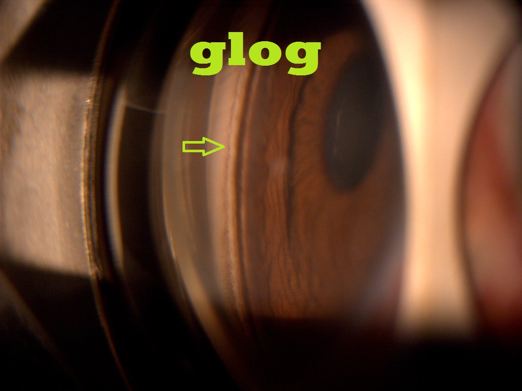
The infant eye, especially the trabecular meshwork, is non‑pigmented. However, with aging, various grades of pigmentation might become visible.
In most cases, pigment dispersion is innocuous and does not cause any pathological changes in the eye.
This condition is known as Primary Pigment Dispersion Syndrome (PPDS). It is characterized by pigment deposition throughout the anterior segment.
The common sites of pigment deposition are: the corneal endothelium, iris surface, lens zonules and capsules, as well as the anterior chamber angle.
PPDS can convert to open angle glaucoma over time. Thus, it is important to monitor such cases carefully.
-------------------------------------------------------------------------------------------------------------------
CAUSES OF TRABECULAR PIGMENTATION
1. P seudoexfoliation & pigment dispersion2. I ritis
3. G laucoma (following acute angle closure)
4. M elanosis of angles (oculodermal melanosis)
5. E ndocrine (diabetes & Addison's disease)
6. N evus (Cogan-Reese syndrome)
7. T rauma
---------------------------------------------------------------------------------------------------------------------
We recently reported about a patient who also had peripheral retinal pigmentary abnormalities. Please check link:
http://www.ncbi.nlm.nih.gov/pmc/articles/PMC4860978/


No comments:
Post a Comment