Friday, January 26, 2018

SWEPT-SOURCE OCT
(UPDATED)
 

“Swept source” refers to the type of laser incorporated in “Swept-source OCTs”. Instead of the super-luminescent diode laser typically seen in conventional spectral domain OCTs (SD-OCTs), swept-source OCT (SS-OCT) uses a short-cavity swept laser. Although the swept source laser has a wavelength centered around 1µ, the laser actually changes as it sweeps across a narrow band of wavelengths with each scan. Like SD-OCT, SS-OCT has a fixed reference arm, but it does not use a spectroscope due to the tunable laser. Instead, a complementary metal oxide semi-conductor camera is employed, along with 2 fast parallel photodiode detectors. Thus, extremely high scanning speeds of 100,000 A-scans per second can be obtained. SS-OCT also has a high axial resolution of just 5µ and an improved signal-to-noise ratio.


Advantages of SS-OCT=

  • 1.       High imaging speed: This allows high resolution images to be obtained while reducing the negative effect of patient’s eye movements on scan quality.
  • 2.      It uses an invisible light which is less distracting to patients, compared to the visible light used in SD-OCT.
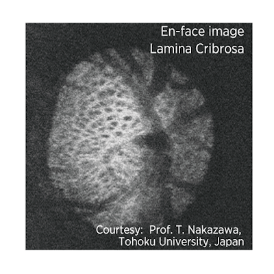
  • 3.      The long wavelength and swept-source technology provide the ability to obtain clear images of deep ocular structures such as choroid and lamina cribrosa which is the probable site of axonal damage in glaucoma.
  • 4.      Imaging of deep structures is possible as the long wavelength of SS-OCT is less subject to light scatter by the retinal pigment epithelium (RPE). There is less light scattering by lens opacities, therefore, SS-OCT can provide clearer images in patients with cataracts, compared to conventional OCT.
  • 5.      SS-OCT provides uniform sensitivity over the entire scan window, which enables the vitreous, retina and deep ocular structures to be visualized in a single scan. In comparison, conventional SD-OCT does not have the same capability and suffers a drop off in sensitivity with changing scan depth.
  • 6.      Placement of a peripapillary circle is not required with the wide-angle scan. In eyes with an atypical optic disc configuration, such as those with tilted optic discs or extensive areas of peripapillary atrophy, placement of the peripapillary circle can be challenging due to difficulties in delineating the optic disc margins.
  • 7.      The SS-OCT is less susceptible to artifacts that may affect the peripapillary circle measurements, such as those produced by floaters, localized scars or extensive peripapillary atrophy extending to the region of the circle.
  • 8.      Due to segmentation software incorporated in the machine, segmentation of the retinal ganglion cell layer (RGC) and inner plexiform layer (IPL) thicknesses across the entire 12x9mm scan is possible. It may provide a means for direct single-scan structure-structure comparisons of peripapillary and macular retinal layers.

Topcon’s Deep Range Imaging OCT-1 (Atlantis) can perform a wide-field scan covering a 12x9mm area of the posterior pole. Therefore, the disc and macula can be evaluated in a single scan. The DRI-OCT uses a center wavelength of 1050µ and a sweeping range of approximately 100nm, compared to the fixed 850nm wavelength typical of SD-OCT. The instrument uses 2 parallel photodetectors to achieve a scan rate of 100,000 A-scans per second compared to 40,000 A-scans per second scanning rate typical of SD-OCT.
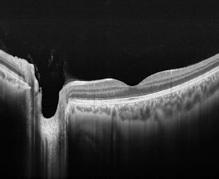
Optic nerve head image obtained by DRI-OCT1


SS-OCT also incorporates automated segmentation software which allows identification of 7 different retinal layers. It is therefore possible to image the circumpapillary retinal nerve fiber layer (RNFL) and macular ganglion cell layer (GCL) using the same scan. The segmentation software also identifies the internal limiting membrane (ILM), IPL, inner segment-outer segment junction (IS/OS), retinal pigment epithelium (RPE), Bruch’s membrane and choroid. Multiple thickness maps can then be generated. 

Possible applications of SS-OCT in glaucoma include: disease detection; identification of novel risk factors; improving the understanding of disease mechanisms. 

In a study conducted by Yang et al, the diagnostic ability of both the wide-angle and peripapillary RNFL thickness measured with SS-OCT were similar to that of peripapillary RNFL thickness measurements obtained with SD-OCT. The average global RNFL thickness measurements acquired by SS-OCT wide-angle scans were thinner than that for peripapillary RNFL scans. This is assumed to be due to more axons in the peripapillary area compared to other areas scanned by wide-angle protocols. However, compared to SD-OCT, SS-OCT had a faster image acquisition rate.


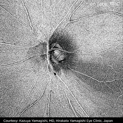
Other studies are being conducted to use SS-OCT in deeper assessment of the lamina cribrosa. SS-OCT is able to scan deeper into the optic nerve head, overcoming image acquisition difficulties due to overlying blood vessels and tissues. This could give us a new perspective on glaucomatous changes in the optic nerve head.


Taken from the article by Yong et al, the above image shows representative swept-source optical coherence tomography (SS-OCT) B-scans of optic discs in high-tension glaucoma (HTG), normal-tension glaucoma (NTG), and healthy eyes.

Horizontal (A, C, E) and vertical (B, D, F) optic disc scans of HTG (A, B), NTG (C, D) and healthy eye (E, F). The image delineated with yellow guidelines is the same as that depicted to the left. The area shaded with yellow depicts the degree of posterior bowing of the lamina cribrosa (LC) according to the level of anterior laminar insertion depth (white solid line). (A, B) Optic disc scans of 65-year-old male with primary open-angle glaucoma (POAG). His baseline intraocular pressure (IOP) was 45 mmHg, and his IOP at examination was 11 mmHg. The overall anterior laminar insertion depth (ALID) was 381.8 μm, the overall mean LC depth (mLCD) was 484.4 μm, and the overall LC curvature index was 102.7 μm. (C, D) Optic disc scans of 65-year-old male with POAG. His baseline IOP was 18 mmHg, and his IOP at examination was 13 mmHg. The ALID was 290.3 μm, the mLCD was 359.9 μm, and the overall LC curvature index was 69.6 μm. (E, F) Optic disc scans of healthy 46-year-old male. His IOP at examination was 13 mmHg. The ALID was 152.6 μm, the mLCD was 146.9 μm, and the overall LC curvature index was –5.7 μm.  

http://www.topcon.co.jp/en/eyecare/products/product/diagnostic/oct/DRI_OCT-1_E.html 

IMAGES RECEIVED FROM TOPCON CORPORATION, JAPAN


Wednesday, January 24, 2018

GLAUCOMA UPDATE: SSY ENGINE

A glaucoma update was organized by Allergan at Le Meridian Hotel, Kuala Lumpur, Malaysia on 20th January 2018. The  highlight of the update was the presence of Dr Anders Heijl from Sweden. A name who requires no introduction, Dr Heijl has worked extensively on visual fields: developing SITA for the Humphrey perimeter, the Visual Field Index (VFI) and now the revolutionary SSY engine to determine target intra-ocular pressure (IOP).

Dr Anders Heijl and myself


The update started with a talk on "Glaucoma through the eyes of patients" by Dr Lee Ming Yueh. This was followed by a presentation by Dr Aziz Husni on "Use of structure and function in clinical decision making in glaucoma".


Dr Lee Ming Yueh

Dr Aziz Husni


After a break, Dr Heijl spoke on "Modern Glaucoma Management in Europe".

Dr Anders Heijl

Two things stood out in his talk:

(1) His team conducted a study in Sweden to assess the "Lifetime risk and duration of blindness in patients with manifest open-angle glaucoma (OAG)". According to the study, published in "Ophthalmology" journal, it was found that at the time of death 42.2% of the glaucoma patients were blind in one eye and 16.4% were blind in both eyes. This means that nearly half of the patients being treated for glaucoma ultimately ended up being blind atleast in 1 eye.

Thus, if we extrapolate this data world-wide, it would indicate that the number of patients blind from glaucoma should be much higher compared to epidemiologic studies conducted by others. This would be an eye opener for ophthalmologists, administrators and NGOs. This data needs to be brought to the notice of those who allocate funds for glaucoma care.


(2) Patients in Dr Heijl's study, as well as some other studies, had apparently well controlled IOP. This shows that there are other factors apart from IOP which can cause optic nerve damage and lead to blindness.

So far most of our efforts have been focused on reducing IOP. These include pharmacologic, laser, surgical and other means such as MIGS and Glaucoma Drainage Devices. Not much is being done to protect the nerve (neuroprotection), investigating the role played by other causes of glaucomatous optic atrophy (vascular, biochemical etc) and neuro-regeneration following optic nerve damage.

This lop-sided focus on reducing only IOP is contributing to a quantum jump in blindness from this disease. 

Dr Heijl's second talk was titled "SSY engine in practice". 


The SSY engine is an app to be used on computers and tablets. Like the glaucoma risk calculator, the app is a simple calculating algorithm. It uses data on measured rate-of-progression, current field status, patient's current age and at diagnosis, as well as measured IOP values. The user can simulate different future rates of progression and see future IOP levels associated with those rates. This helps in better target IOP settings depending on the expected life-span of the individual.


Dr Heijl explaining the SSY engine
The app is to be used when the measured rates of progression are available and the treating ophthalmologist is not content with the results. So, this app cannot be used at the time of diagnosis as we do not know the rate of progression at that time and would be useful after 2-3 years of diagnosis when rate of progression is more evident. 

The program was followed by a sumptuous dinner.

Thursday, January 11, 2018

SUSTAINED RELEASE MEDICATIONS IN GLAUCOMA


Compliance to anti-glaucoma medications remains a major limiting factor in the management of glaucoma. With old age come physical difficulties in instilling medications, dementia leads to forgetfulness of the doses and the financial constraints in buying medications indefinitely puts a strain on the pocket of old pensioners. In order to overcome the factor of non-compliance to anti-glaucoma medications, sustained release drug delivery systems are being investigated. 


These long-acting or enhanced delivery systems can be broadly divided into EXTERNAL and INTERNAL platforms, depending upon their location with respect to the coats of the eyeball.


EXTERNAL PLATFORMS:


1. PUNCTAL PLUGS= Drug-infused plugs which can be inserted into the punctum are undergoing clinical trials. These include the Ocular Therapeutix’s Travoprost (OTX-TP) and the Mati Therapeutic’s Latanoprost plugs. The former is cylindrical while the latter is arrow shaped to prevent inadvertent slippage into the canaliculi. The OTX-TP is expected to provide sustained drug delivery for 2-3 months. 


OTX-TP

Mati Therapeutics punctal plug

With the Mati Therapeutic’s plug, IOP reportedly dropped by 6 mmHg after 1 week. 


Side effects of plugs include falling out or intra-canalicular migration, local skin darkening near the medial canthus and epiphora due to the plug blocking the punctum.


2. RINGS= A ring, containing Bimatoprost, which fits into the superior and inferior fornices is being developed by Allergan. The retention rate of the ring is reportedly around 90% at 6 months and the efficacy lasts for 4-6 months. The loss of efficacy is presumably due to the elution of the drug with time and the blockage of receptors due to continuous exposure to the drug. When the ring was re-inserted after 6 months of initial use, the same effectiveness as in the first insertion was not seen, probably due to the down-regulation of the receptors.


3. PERIOCULAR INJECTION= A subconjunctival bioerodable pellet containing Latanoprost is being developed. Known as the Durasert, the 3 mm implant is injected using a 27-gauge needle. Pfizer and pSivida are conducting a Phase I/II safety and efficacy trial of the Durasert.


4. SUBCONJUNCTIVAL INJECTION= Natarajan JV and colleagues reported Latanoprost incorporated into LUVs (Large Unilamellar Vesicles) derived from the liposome of DPPC (di-palmitoyl-phosphatidyl-choline) by a film hydration technique, and injected subconjunctivally. The IOP lowering efficacy of this vehicle was sustained upto 50 days.


https://www.sciencedirect.com/science/article/pii/S0168365914003320 


5. CONTACT LENSES= (a) Latanoprost eluting low dose contact lenses (CLLO) and high dose contact lenses (CLHI) have been produced by encapsulating a thin latanoprost-polymer film within the methafilcon hydrogel lathed into a contact lens. As reported by Ciolino and colleagues in Ophthalmology journal, CLLO reduced IOP by 6.3+/-1.0 (at day 3), 6.7+/- 0.3 (at day 5) and 6.7+/-0.3 (at day 8). The CLHI reduced IOP by 10.5+/-1.4 (at day 3), 11.1+/-4.0 (day 5) and 10.0+/-2.5 (day 8). Topical Latanoprost reduced IOP by 5.4+/-1.0 mmHg on day 3 and 6.6+/-1.3 mmHg on day 5. Thus, sustained delivery of Latanoprost by contact lens was found to be atleast as effective as the topical application. (b) Hiratani and Alvarez-Lorenzo have reported the usage of soft contact lenses consisting of polymers of N,N-diethylacrylamid and methacrylic acid which appear to deliver timolol for approximately 24 hours.

 https://www.ncbi.nlm.nih.gov/pubmed/27586444

https://www.ncbi.nlm.nih.gov/pubmed/12363448 


6. TOPICAL OPHTHALMIC DRUG DELIVERY DEVICE (TODDD) = This device developed by Amorphex Therapeutics has combined polymers which allow sustained release of Timolol. The device is put under the upperlid, where it floats over the tear film. It has a corneal relief curve which prevents it from riding over onto the cornea. IOP was found to be reduced by 16-22%. The company is planning Phase I trials for the TODDD. The device will  probably be effective for 90 days.


INTERNAL PLATFORMS:


1. Allergan’s bimatoprost sustained-release implant, which can be injected into the anterior chamber, is undergoing Phase III trials. In Phase II trials it reduced IOP in 92% patients at 4 months and in 71% at 6 months. The pressure lowering was by 8-10 mmHg at week 2, but by week 26 the IOP lowering was by 6-8 mmHg. The advantage of such implants is that there is no risk of dislodgement. 


2. ENV515 (Envisia): This intracameral implant uses Travoprost. It uses a biodegradable polymer drug delivery system. In Phase I trials IOP reduced an average of 35% (6.4+/-0.6 mmHg) over 8 months, but returned to baseline at 9 months. In Phase IIa clinical trials IOP was reduced by 28% (6.7 mmHg) at day 25, which was comparable with instillation of Travatan Z once daily in the other eye. The only side effect noted was transient hyperemia.


3. GrayBug is developing a microparticle technology originally created at the Wilmer Eye Institute. 3 possible agents: a single IOP lowering compound; a dual-action IOP-lowering compound and another IOP-lowering agent with neuroprotective properties, are being developed. The agents will be injected intravitreally or subconjunctivally. The implant would be absorbed over a 6-month period.

http://graybug.com/pipeline/gb-201-202-203-glaucoma/ 


4. Clearside Biomedical and Santen are developing a supraciliary drug delivery system using Clearsides’ microinjector and Santen’s sustained release formulations. Sulprostone and Brimonidine were tested with the delivery system and decreased IOP significantly compared to their topical counterparts. In rabbit eyes, a single injection of Brimonidine reduced IOP by 6 mmHg. However, the effect tapered off after a month.

https://www.businesswire.com/news/home/20150504005229/en/Clearside-Biomedical-Santen-Announce-Research-Collaboration-Glaucoma 


5. Ohr Pharmaceuticals is developing injectable micro- or nano-articles using Latanoprost. The particles can potentially be injected within or around the eye. Adnexal adverse effects do not occur with this agent. However, there is risk of toxicity, damage to intraocular structures and need for lifelong procedures to ensure sustained lowering of IOP.

6. Icon Bioscience is developing a biodegradable injectable implant to deliver Latanoprost. The IBI-60089 is injected intracamerally through a 30G needle.

 http://iconbioscience.com/pipeline/


7. Euclid Systems is developing 2 collagen based systems to provide sustained release of Latanoprost. The first is an injectable insitu gelling collagen solution and the second a 2mm x 4mm collagen wafer implanted over the sclera. The latter has shown the release of Latanoprost to last upto 180 days.

8. Replenish has developed the Ophthalmic Micropump which is implanted over the sclera. The wireless connected and programmable system dispenses nano-liter sized doses of drugs, which can last upto 12 months. Once the medication is exhausted, the device can be refilled with a 31G needle. 


Thank you for visiting the "glog ".

LIGHT-ACTIVATED LIPOSOMES FOR GLAUCOMA

  Biomedical researchers at Binghamton University in the USA, have developed a mechanism for drug-carrying liposomes that can be activated i...