Tuesday, June 30, 2020

DYNAMIC CONTOUR TONOMETRY


INTRODUCTION:

Corneal abnormalities are known to affect tonometry measurements. These abnormalities can occur due to diseases or after surgical procedures such as keratoplasty or scleral buckling. A number of methods have been described to overcome them. In the Goldmann Applanation Tonometer (GAT), taking 3 readings and then taking a mean or taking the reading in one axis and then rotating the head by 900 have been suggested. Some instruments claim to offer better measurements of intra-ocular pressure (IOP) by overcoming corneal factors such as hysteresis. The Ocular Response Analyzer (ORA) is one such instrument. A recent advance is the dynamic contour tonometer.

The PASCAL Dynamic Contour Tonometer (DCT) from Ziemer is a slitlamp mounted tonometer which claims to provide true IOP, independent of interindividual variations in corneal properties and biomechanics confirmed with intracameral measurements. It also apparently offers the highest repeatability for a refined IOP and glaucoma progression analysis, and determines Ocular Pulse Amplitude (OPA), which has been correlated to glaucoma risk.


The DCT does not require fluorescein, only local anesthetic is instilled. The device is centered in the middle of cornea and the device advanced forward until the lever arm is upright. Fine adjustments are made through the slit-lamp such that the contact area (black rim) is centered nicely. The Q-indicator-tone indicates proper measurement of IOP. The device can be connected to wireless printer or PC and is EMR compatible. It also has automatic self calibration, therefore, no additional calibration steps are needed. The biggest advantage in the present scenario of COVID-19 pandemic is the low risk of cross infection due to sterile sensor caps.

DCT PRINCIPLE:

Hartmut Kanngiesser, PhD, and Yves Robert, MD, (at the Swiss Federal Institute of Technology and at the University Hospital of Zurich, Switzerland, respectively) developed the theoretical basis and the clinical proof of concept for a radically different methodology, which they termed dynamic contour tonometry (DCT). Their device's contoured tonometer tip has a concave surface, which touches but allows the cornea to assume a shape (close to its steady-state shape). In such a state no tangential and bending forces act within the area of the cornea touching the tip. If the apex of the cornea is tension-free, the pressure acting on both of its sides (inside and outside) must be exactly equal. The presence of a miniaturized piezoelectric pressure sensor flush inside the surface contour of the tonometer's tip allows practitioners to measure the “extra-ocular pressure” at the corneal apex in order to obtain a direct reading of the IOP at the opposite side of the cornea.

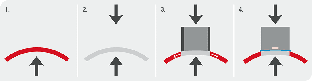
Due to IOP there is pressure on the cornea from the inside. However, due to the collagen matrix in the cornea, the corneal shape is maintained. The cornea is regarded as tension-free when the internal and external pressure across the cornea is equal. This is called Pascal’s Law. If the pressure in a small part of the cornea is equalized, the center becomes tension-free and flattens. This shape of the cornea is called the DCT-shape and the tonometer tip fits nicely in this shape. In such a tension-free state of the cornea the device measures true IOP, as if there is no cornea.

EFFECTIVENESS OF DCT:

Detailed mechanical analysis has demonstrated that variations in any corneal properties over a wide range of values does not influence this pressure measurement, and it showed that even the amount of appositional force applied to hold the tip in place does not affect the pressure reading.

Investigators analyzed tonometer data obtained with DCT, GAT, and pneumatonometry by plotting them against corneal thickness in healthy volunteers. Data from both GAT and pneumatonometry exhibited a significant correlation with corneal thickness; the measurements increased on average by 0.35 mm Hg per 10 µm of corneal thickness. In marked contrast, the DCT data were flat across the spectrum of corneal thickness readings, a finding that suggests that this tonometer alone functions independently of corneal thickness.

The chosen contour of the DCT tip will furnish correct measurements (defined as measurements with a systematic error of less than 0.5 mm Hg) for corneas with a radius of between approximately 5.5 and 9.2 mm and a corneal thickness ranging from 300 to 700 µm. Outside these limits, the systematic error will gradually increase. It is therefore important to note that some corneas will not satisfy the requirements for correct IOP measurements with DCT. Most nonpathologic corneas will be well within the scope of what DCT can handle correctly, however.

In a study performed in vivo, Andreas Boehm compared IOP in the anterior chamber with Pascal measurements prior to cataract surgery. (Boehm et al, 2008) This study demonstrated that DCT values were reliably within one millimeter of actual manometric IOP. In a more recent comparison between GAT and DCT IOP, Kotecha et al showed that DCT was more precise and reliable than GAT. (Kotecha et al, 2010).


Saturday, June 6, 2020

FOURTH ANNIVERSARY OF THE GLOG

At the start of the year 2020, I wrote: "Perhaps this year is the most anticipated period in the lives of ophthalmologists". Our expectations at that time were sky-high. My dream was to see this year a stimulus for growth. Halfway down 2020, this period seems to have turned into an uncomfortable nightmare. Whatever optimism we had at the start of the year has lost steam. Now, it's a struggle to survive physically, economically, socially and emotionally. That's all thanks to a tiny Coronavirus.

The COVID-19 pandemic has affected the lives of humanity in general and patients/health care workers in particular. The world's population has come face-to-face with the biggest calamity in recent history. Lives have been lost from the disease itself and incidents surrounding the pandemic. This has been the worst calamity to affect our social fabric.

For the first time we posted something apart from glaucoma on The Glog. Some of the posts have focused on COVID. And, one article was shared on the touchOPHTHALMOLOGY website in its Insight section.

The Glog tried its best during the last one year to present glaucoma-related topics in simple, easily understandable posts. The most memorable part was to have The Flammer Series. In those 4 articles, we have attempted to show a small glimpse of the profound genius of Prof Josef Flammer. In my opinion Prof Flammer has done such immense work on glaucoma that he deserves to be put on the pedestal of all-time greats of glaucoma. A humble and gentle being he has not advertised himself, unlike many other loud-mouthed individuals who appear to present themselves as the greatest thinkers since Socrates and Plato. No words are enough to acknowledge the contribution of Prof Flammer to glaucoma research.

Last year we had also focused on Artificial Intelligence (AI) in glaucoma. And, just last week I received a survey from the Asia Pacific Ocular Imaging Society to look into AI practices. So, surely AI is picking up as predicted.

Another leap forward for The Glog was the large number of articles from students. They are the future of our society and I hope these young men and women will carry the responsibility to attain greater heights in healthcare.

I cannot thank enough the visitors to the site. For me, there’s no other stimulus to work than the knowledge that my efforts are going to a wider audience. At the time of posting this article, there have been 45,282 page views to The Glog. My humble appreciation for patronizing The Glog over all these years. Our free-to-access blog is an amateurish attempt to bring glaucoma-related knowledge to a wide audience.

The Glog continues to explore new ways to present Ophthalmology. We are working on some innovative ideas to improve our presentation. Do keep checking here for the updates.

Your suggestions to improve The Glog are most welcome.

I wish each and everyone a safe, healthy and happy future.

Take care

Shoeb


Tuesday, June 2, 2020

iStent MIGS device

INTRODUCTION

The iStent is the smallest device ever implanted in the human body. This minimally invasive glaucoma surgery (MIGS) device is devised to bypass the obstructed trabecular meshwork (TM) and allow flow of aqueous from the anterior chamber into the Schlemm’s canal. The instrument was devised by the Glaukos company in the USA and one of the earliest published report was by Spiegel et al. in 2007. The first generation iStent was 1 mm in length and 0.3 mm in height, making it the smallest device approved for implantation by FDA in 2012. This stent is shaped like a snorkel with retention ridges. It is made from an implant grade, heparin-coated titanium alloy and is inserted in the TM manually. The stent is implanted by an ab interno approach guided by gonioscopy.


Subsequently, in 2018 the company came up with the iStent Inject. This device comes with two stents within the injector, as opposed to the first-generation device that had only one stent. Similarly, it is also composed of implant-grade titanium and is 360 μm in length and 230 μm in width, which is one-third the size of the first-generation iStent. Unlike the snorkel shaped first generation iStent, the second generation stent contains a central inlet and multiple outlets designed to provide multidirectional flow through Schlemm’s canal.


Studies regarding these devices have usually been conducted utilizing combined cataract surgery with iStent implantation. Most of the studies have shown significant reduction in intra-ocular pressure (IOP) and medication usage. One of the major studies regarding this device has been the US iStent Study Group. The study compared combined cataract surgery with a single iStent implantation versus phacoemulsification alone. 70% patients in the combined group achieved IOP <22 mmHg without medications compared to 50% in the phacoemulsification alone group (p <0.001). At 12 months, the number of patients requiring medications in the combined group was 15% compared to 35% in the phacoemulsification alone group (p=0.001). These effects were also found in a follow-up study conducted at 24 months.

Studies on solo iStent implantation have also demonstrated >20% IOP lowering until 24-months of follow up. Studies have compared 1,2 and 3 iStent implantations. It was found that 2 and 3 iStent implantation led to nearly 41% IOP reduction, compared to 31% by single stent use.

Therefore, it can be concluded that iStent is a useful trabecular bypass MIGS device which can be effective in IOP lowering as a solo or combined procedure.

SURGICAL TECHNIQUE

The surgical steps for iStent implantation are available in detail elsewhere (See link). Here, the steps are given in brief. In the first generation iStent, the anterior chamber is filled with viscoelastic. A temporal clear-corneal incision is made. If combined with cataract surgery the same incision as the one for phacoemulsification can be used. The iStent on its injector is pushed into the AC and using a gonioscope to guide, is directed towards the nasal angle in such a way that the snorkel is facing downwards. The stent enters the TM and with a sideways motion of the hand the device is pushed deep inside. Subsequently, the inserter button is pressed, allowing the device to settle into the position. Small adjustments are made by tapping the end of the snorkel by the inserter. Finally, the inserter is taken out of the eye and the AC cleared of the viscoelastic.

The iStent Inject comes with 2 pre-loaded implants. The surgeon enters the eye from the temporal clear-corneal incision and using the gonioprism reaches the TM. Then the device is brought into approximation at the desired position and the button pressed without any side to side movement of the hand to allow the implant to be delivered into the desired spot. The second stent is then implanted similarly 2-3 clock hours away.

https://www.youtube.com/watch?v=LCDE0NB6S2Y

COMPLICATIONS

Most studies did not report any complication from the iStent per se and most were attributed to cataract surgery. A few studies have reported on stent occlusion or malposition occurring in 2.6-18% patients. The new iStent Inject provides multiple openings to minimize the possibility of occlusion. Mild bleeding during implantation of the iStent is common, but usually resolves spontaneously.

A study was conducted in Canada regarding cost effectiveness of the device.  The potential savings of the iStent compared to a 2-drug regimen over a 6-year period was CA$1272 and CA$2124 when using 3 drugs.

CONCLUSION

 The iStent is a useful addition to our choice of MIGS devices. It is technically easy to use, efficient and safe.


HELICOBACTER PYLORI ASSOCIATED GLAUCOMA

Helicobacter pylori (H. pylori) is a spiral-shaped and gram-negative micro-organism that affects the epithelial mucosa of the stomach. In t...