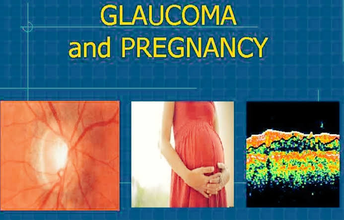
PREGNANCY AND GLAUCOMA
GUEST AUTHOR
SANIA KHAN
AJMAL KHAN TIBBIYA COLLEGE
ALIGARH, INDIA

INTRODUCTION
Although
glaucoma is a disease of the older population, but it may sometimes affect
women of childbearing age.
Glaucoma
is rarely diagnosed during pregnancy; occasionally patients with preexisting
glaucoma become pregnant, however.
Management
of glaucoma during pregnancy is a challenge due to side effects and teratogenic
effects of medications, as well as the risk of surgery on the mother and fetus
has to be considered.
This
post takes a look at the features of glaucoma during pregnancy and it's
management.
IOP AND PREGNANCY
Intraocular pressure (IOP)
is decreased by 10% in women during pregnancy with normal eyes.
In
glaucomatous pregnant women with Ocular hypertension (OHT), there is significant
decrease in IOP as compared to normal pregnant women without OHT.
IOP
decline in non-glaucomatous pregnant women occurs between 12-18 weeks of
pregnancy. While in women with OHT this decline occurs between 24-30 weeks.
The
ocular hypotensive effect of late pregnancy is significantly greater in
multigravidae than in primigravida due to an effect of the increased stress and
discomfort felt by the primigravidae.
This
stress can induce the release of large quantities of epinephrine and
norepinephrine, leading to a higher IOP. However, the effect of psychological
stress on IOP has never been proven.
The
exact cause of lowered IOP during pregnancy in healthy women is unknown.
It
is proposed that increased uveoscleral outflow due to changes in hormonal levels.
Estrogen,
relaxin, progesterone and beta-chorionic gonadotropin are the suspect hormones.
Acidosis
and decreased aqueous production are also proposed as a cause of lowered IOP
but aqueous flow rate has been found to be consistent during pregnancy.
Decreased
episcleral pressure during pregnancy is also proposed as the mechanism for
lowered IOP.
IOP
measurement errors due to reduced corneo-scleral rigidity, making tonometry
readings falsely low, is another factor.
The
physiologic changes of pregnancy are so numerous and complex that it is very
difficult to reach any definite conclusion.
The
highest drop of IOP in the third trimester of pregnancy was 2.7 mmHg as
compared to non-pregnant women.
Reduced
diurnal variation of IOP as well as lowered IOP in the third trimester have a
protective effect in pregnant patients with glaucoma.
Pregnant
women may avoid treatment of glaucoma to protect the fetus, thereby increasing
non-compliance.
CORNEA AND PREGNANCY
A
measurable, but slight increase in corneal thickness has been found in pregnant
women.
The
central corneal thickness was increased 16 microns (p=0.1) compared to the
control eyes. This increase occurs due to water retention.
The
lower IOP in pregnant patients did not correlate with corneal thickness.
VISUAL FIELDS IN
PREGNANCY
The
visual field changes during pregnancy include bitemporal contraction,
concentric contraction and enlargement of the blind spot.
Proposed
mechanisms for these visual field changes are diverse.
MRI
studies show that the size of pituitary gland is increased about 120% during a
normal pregnancy, so it is believed that the pituitary impinges on the optic
chiasm causing the temporal hemifield deficit, the most common visual field
defect in glaucoma is in the nasal hemifield.
The
rarity of glaucomatous visual field defect in the temporal visual field and
reversibility of any kind of VF defect after pregnancy are important features
to distinguish between glaucomatous and pregnancy induced VF defects.
RETROBULBAR BLOOD
FLOW IN PREGNANCY
Altered
retrobulbar blood flow plays an important role in the pathogenesis of open
angle glaucoma.
Vascular
dysregulation in susceptible patients, independent of atherosclerosis is
thought to reduce ocular blood flow.
Alterations
in hormonal status leading to changes in CVS are also implicated.
The
influence of menopause and post menopausal hormone supplementation on the
incidence of coronary artery disease in women has been explained in various
studies and this seems to be linked to the "estrogen vasodilator
activity".
Blood
flow velocities in the retrobulbar arteries are significantly increased by
estradiol.
Ocular
Blood Flow changes may occur due to physiologic and progressive increase of estrogen
secretion during pregnancy.
There
may be significant increase in pulse ocular blood flow in pregnant women as
compared to non-pregnant women.
The
pulse Ocular Blood Flow is 500 ml/min in the first quarter of pregnancy and 600
ml/min in the second quarter greater than in non-pregnant women.
Resistive
index and pulsality index are inversely proportional to the gestational age.
During
pregnancy there is an increase in estrogen that induces endothelial-dependent
vasodilation in several tissues and maybe cause of increased ocular blood flow.
Intranasal
administration of 17-beta estradiol increased the peak systolic and end
diastolic velocities of the Central retinal artery without any significant
difference in ophthalmic artery flow velocities or pulsality and resistive
indices.
Glaucomatous
and non-glaucomatous postmenopausal women who received hormone therapy may show
a significant decrease in ophthalmic artery pulsatile index.
The
Ophthalmic artery mean pulsality index decreases significantly more in
non-glaucomatous women than in glaucomatous women (-43% vs. 28%; p=0.001).
Patients
with lower retinal blood flow velocities may show higher rates of progression
of glaucomatous damage.
In
some women with increased retrobulbar blood flow and lower IOP during
pregnancy, the protective effect of pregnancy on glaucoma progression is
possible.
LABOR IN GLAUCOMATOUS
PATIENTS
Angle-closure
glaucoma may be precipitated during labor in women with narrow angles.
The
mean IOP increased by 1.4 mmHg during the course of labor then decreased by 3.0
mmHg immediately after delivery. The IOP returned to pre-labor levels in all
patients by 72 hours after delivery.
Oxytocin
levels increase during labor which leads to capillary constriction and decrease
in aqueous outflow but there is no evidence linking oxytocin to IOP.
Valsalva
maneuvers also increase IOP during labor but their effect on progression of
glaucoma is not proven.
Some
studies found a higher risk for glaucoma progression in patients with low blood
pressure. Transient hypotensive shock may also lead to glaucoma-like optic
nerve and VF changes.
Large
amount of blood loss during labor leads to transient hypotension and increased
risk for glaucoma progression.


No comments:
Post a Comment