Wednesday, March 20, 2019

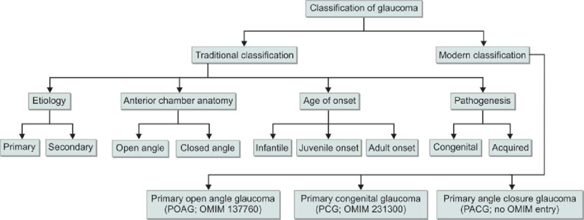
CLASSIFICATION OF GLAUCOMAS


The classification of glaucomas has seen a progressive change as our understanding of this condition has evolved. The anatomic, gonioscopic, biochemical, molecular and genetic basis for the classification of glaucomas has been utilized, each having its own pros and cons. With the advent of new instruments to diagnose glaucoma, classifications have also been created based on the techniques utilized. Thus, the classifications of glaucoma include those based on:

-Etiology
-Mechanism
-Staging
-Optic nerve appearance
-Visual field damage
-Standard HRT parameters by bagging classification trees
-Automated classification of glaucoma stages using higher order cumulant features
-Texture features using neural networks

Unfortunately, none of the classifications have been satisfactory in their attributes to describe glaucoma. This is not unexpected, since there are different mechanisms of the disease and multifactorial pathogenetic factors at work in different individuals. Presently, the classification of glaucomas based on etiology and mechanism is still applied in clinical practice, having stood the test of time over the years.

Etiologic Classification= This is based on the underlying disorder causing alteration in aqueous inflow/outflow (Aqueous humor dynamics) or Retinal Ganglion Cell (RGC)/Optic nerve damage. 
Mechanistic Classification= This is based on specific alteration in the anterior chamber (AC) angle that causes intra-ocular pressure (IOP) to rise.

These classifications have incorrectly been based on our focus on elevated IOP as the major risk factor for the development of glaucoma, excluding other factors such as vascular, genetic or biochemical mechanisms among others.

CLASSIFICATION BASED ON ETIOLOGY 

Based on the etiology, glaucomas have been divided into primary and secondary. The primary glaucomas are assumed to have the initial events leading to outflow obstruction and IOP elevation primarily in the AC angle or conventional outflow pathway. These glaucomas are not associated with known ocular or systemic disorders which could impede aqueous outflow. They are usually bilateral and probably have a genetic basis. From a therapeutic standpoint it is essential to differentiate open angle glaucoma from closed-angle glaucoma.

On the other hand, secondary glaucomas are regarded as such because of a “partial understanding of the underlying, predisposing ocular or systemic events” [Bruce Shields]. These are usually asymmetric or unilateral. While some may have a genetic basis, others are acquired. As the concepts regarding the underlying causes of the glaucomas continue to develop, the primary and secondary classifications have become increasingly artificial and inadequate.

Classification of childhood glaucomas, especially those associated with developmental anomalies of the anterior chamber angle have been dogged by overlapping and variably defined nomenclatures which frequently denotes the age of onset rather than the underlying mechanism for the glaucoma. 

Bruce Shields has recommended replacing traditional concepts with a new scheme that provides a “better working foundation for the concepts of mechanism, diagnosis and therapy that will shape the management of glaucomas for the foreseeable future”. He has classified glaucomas based on staging. According to him, glaucomas can be considered to consist of 5 stages:

Stage I: Initiating events
Stage II: Structural alterations
Stage III: Functional alterations
Stage IV: RGC and ON damage
Stage V: Visual loss


The initiating events (Stage I) are speculated to have a genetic basis. Structural changes may start occurring in the RGCs or optic nerve head (ONH), as a result of alterations in proteins in these regions. These structural alterations (Stage II) could be subtle tissue changes in the blood vessels supplying the ONH or in supportive elements of the lamina cribrosa. Or they could act through mechanisms as yet to be understood. Structural changes may lead to functional alterations (Stage III) such as reduced axonal conduction, vascular perfusion to axons in the ONH or a progressive deformity of the lamina cribrosa that may lead (alone or in conjunction with a relative IOP elevation) to glaucomatous optic neuropathy (Stage IV), which gets reflected in subsequent VF changes (Stage V).

Traditionally, glaucomas have been divided into open and closed angle.

Chronic open angle glaucoma: This is characterized by optic nerve damage in an eye which does not have evidence of angle closure on gonioscopy and there is no identifiable secondary cause. Apparently inherited susceptibilities lead to increased resistance to aqueous outflow and higher vulnerability of the ONH to the level of IOP.

Pupillary block glaucoma: Primary Angle Closure includes asymptomatic individuals with occludable angles who have not had an acute attack, as well as those who had an attack which resolved spontaneously or with treatment prior to the development of any detectable nerve damage. Primary Angle Closure Disease (PACD) has been classified by the International Society for Geographical and Epidemiological Ophthalmology (ISGEO) into:

(1) Primary Angle Closure Suspect (PACS): Such eyes have iridotrabecular contact for atleast 2700 and normal IOP, ONH and VFs.
(2) Primary Angle Closure (PAC): There is iridotrabecular contact for atleast 2700 and raised IOP and/or peripheral anterior synechiae (PAS), but with normal ONH and VFs.
(3) Primary Angle Closure Glaucoma (PACG): There is PAC with evidence of glaucomatous damage in the ONH or VFs.
(4) Acute Angle Closure Crisis: There is periocular or ocular pain, often accompanied by headache, nausea or vomiting, IOP >21 mmHg, circumcorneal congestion, corneal edema, mid-dilated pupil and shallow anterior chamber.

Developmental anomalies of AC Angles:
These represent incomplete development of structures in the conventional aqueous outflow pathway. These anomalies could be inherited or acquired during intra-uterine life and lead to elevation of IOP. In some cases the developmental anomaly is not associated with primary or systemic etiologies and regarded as primary.

Pediatric glaucomas have been classified into the following categories:

(1) Primary Congenital Glaucoma (PCG): Primary congenital glaucoma that occurs at or shortly after birth or glaucoma of any etiology that occurs in the same time frame.
(2) Primary Infantile Glaucoma: It is genetically identical to PCG but presents 1-2 months after birth.
(3) Juvenile Open Angle Glaucoma: There is no ocular enlargement; absent congenital ocular anomalies or syndromes; Open, normal appearing angles; meets the glaucoma definition.
(4) Developmental Glaucoma: This term has been used as a giant waste basket for nearly all childhood glaucomas that are not acquired immediately after birth.

Glaucomas associated with other ocular disorders
This class includes those glaucomas in which the initiating event is an abnormality of the ocular structures such as corneal endothelium, iris, ciliary body, lens, vitreous, retina and so on. Or the initiating event is a definite second ocular pathology such as tumor, hemorrhage, inflammation and so on. Secondary glaucomas are properly considered to represent those eyes in which a second form of ocular pathology has caused IOP to rise above the normal range with consequent ON damage. The second ocular pathological processes causing optic neuropathy may include=
      i.        Neovascularization
     ii.        Uveitic conditions
    iii.        Trauma
   iv.        Lens-related

CLASSIFICATION BASED ON MECHANISM

Elevated IOP is the major risk factor for the development of glaucoma. However, the concept that statistically raised IOP is a defining characteristic of glaucoma has been almost universally discarded. A disadvantage of this mechanistic system is that it ignores the causes unrelated to IOP. Also, many of the glaucomas have more than one mechanism of outflow obstruction at different times in the course of disease. As a result some of the glaucomas must be classified under more than one mechanistic heading. On the plus side, the advantage of this classification is that our understanding of the mechanisms of aqueous outflow obstruction is usually more complete than our knowledge of initiating events. An understanding of the mechanism that leads to aqueous outflow obstruction is important in developing a rationale for controlling the IOP in each form of glaucoma.


Mechanisms of Open Angle Glaucoma=
The elements obstructing aqueous outflow may be located on the anterior chamber side of the trabecular meshwork [TM] (pretrabecular mechanisms); in the TM (trabecular mechanisms) or distal to the meshwork, in the Schlemm’s canal or further along the aqueous drainage system (post trabecular mechanisms).

Angle closure glaucoma mechanisms=
Angle closure mechanisms are the ones which cause apposition of the peripheral iris to the TM or peripheral cornea. The peripheral iris may be pulled (anterior mechanisms) or pushed (posterior mechanisms) into this position. In anterior mechanisms usually a contracting membrane in front of the iris pulls the iris towards the TM/peripheral cornea. It can also be caused by consolidation of inflammatory products in this area.
In posterior mechanisms pressure behind the iris, lens or vitreous causes the peripheral iris to be pushed into the anterior chamber angle. These mechanisms can occur with or without pupillary block. Pupillary block variants include pupillary block glaucoma in which there is apposition of the mid-periphery of the iris and the lens, thus blocking the egress of aqueous anteriorly through the pupil. The peripheral iris balloons in the form of “iris bombe”. The functional apposition in these patients is due to a genetically influenced configuration of the anterior segment of the eye. Such appositions may also be seen in lens-induced mechanisms such as phacomorphic glaucoma or ectopia lentis. Pupillary block can also occur from posterior synechiae. The “pushing” mechanisms can also occur without pupillary block such as ciliary block (malignant glaucoma), lens induced, forward shift of vitreous following lens removal, intraocular tumors, cysts of uveal tract, retrolenticular tissue contraction as in retinopathy of prematurity or persistent fetal vasculature.

Developmental anomalies of the AC Angles=
These represent incomplete development of structures in the conventional aqueous outflow pathway. Examples of these include: congenital glaucoma, Axenfeld-Reiger syndrome, Peter anomaly and iridocorneal adhesions.


Sunday, March 10, 2019

PARAPAPILLARY ATROPHY


The Optic Nerve Head (ONH) is often surrounded by different zones of atrophic-like changes occurring in the retina and choroid. These zones may vary in width, circumference or pigmentation. Since the atrophy usually is adjacent to but does not surround the optic nerve (ON) completely, the term “parapapillary” is preferable to “peripapillary atrophy”. However, these terms are often used interchangeably in literature. 

These parapapillary atrophic changes were first described by Elschnig and Bucklers who termed it “halo glaucomatosus”. Primrose also reported on parapapillary glaucomatous changes. According to him:”Peripapillary halo was present in more than half, often quite early in the disease, and was also present in many fellow eyes as yet free from cupping”. He suggested the peripapillary halo could be a useful diagnostic sign in early glaucoma. 

Parapapillary atrophy (PPA) can be divided into 2 types, based on location and appearance:
Zone beta is located closest to the optic nerve head. There is loss of the retinal pigment epithelium (RPE) and most of the photoreceptors in this area so that the sclera and large choroidal vessels become visible. 

Zone alpha is located circumferentially away from the nerve. In this zone there is irregular arrangement of RPE cells, resulting in both hypo- and hyper-pigmentation. The pathological change is pigmentary disruption of the RPE. It is also called “chorio-pigment-epithelio-retinal atrophy”. 

Zone Beta
Beside the nerve
Choroidal vessels and sclera visible
Zone Alpha
Away from nerve
Hypo/Hyper pigmentation


Jost Jonas was probably the first to describe Zones Alpha and Beta. According to him:”The parapapillary chorioretinal atrophy was divided into 2 zones: Zone “Alpha” was characterized by an irregular hypo- and hyper-pigmentation; and intimated thinning of the chorioretinal tissue layer. It was adjacent to the retina on its outer side and to zone “Beta”, or the parapapillary scleral ring of Elschnig on its inner side. Characteristics of Zone Beta were: marked atrophy of the RPE and choriocapillaris, small grey fields on a whitish background, good visibility of the large choroidal vessels, thinning of the chorioretinal tissues, round bordering to the adjacent Zone Alpha on the peripheral side and to the peripapillary scleral ring on the central side. If both zones are present in the same sector, Zone Beta was always closer to the optic disc than Zone Alpha.”


Jonas etal in their study of chorioretinal atrophy in normal and glaucoma patients reported that PPA as a whole and both zones Alpha and Beta were significantly (p <0.00001) larger and Zone Beta was significantly (p <0.00001) more frequent in the glaucoma group than in the control group. The size and frequency of PPA were significantly correlated (p <0.0001) with the glaucoma stage. 
(Jonas JB, Nguyen XN, Gusek GC etal. Parapapillary chorioretinal atrophy in normal and glaucoma eyes. I Morphometric data. Invest Ophthalmol Vis Sci. 1989;30:908-18.)

Jonas and colleagues in another article have reported that PPA was larger and occurred more often in patients with glaucoma (compared to normal or those with ocular hypertension). PPA enlarged as the neuroretinal rim (NRR) area decreased and showed a spatial correlation to VF loss. (Jonas JB, Fernandez MC, Naumann GO. Glaucomatous parapapillary atrophy. Occurrence and correlations. Arch Ophthalmol. 1992;110(2):214-222) 


Tezel etal in their study of PPA in ocular hypertension (OHT) reported that PPA, higher PPA area-disc area, Zone Beta area-disc area and PPA length-disc circumference ratios at the baseline examination was associated with conversion to glaucoma. Intra-ocular pressure (IOP) (relative risk 1.19), NRR area-disc area ratio (relative risk 0.72) and Zone Beta area-disc area ratio (relative risk 1.32) were found to be associated with the development of optic disc damage, Visual Field (VF) damage or both.
(Tezel G, Kolker AE, Kass MA etal. Parapapillary chorioretinal atrophy in patients with ocular hypertension. I. An evaluation as a predictive factor for the development of glaucomatous damage. Arch Ophthalmol. 1997;115:1503-508)

Tezel etal also reported in a study that PPA was already present in 48 (49%) of 98 eyes diagnosed with OHT and which converted to glaucoma. PPA progression was seen in 25 (10%) of 252 ocular hypertensives who did not develop optic nerve or VF damage.
(Tezel G, Kolker AE, Wax MB etal. Parapapillary chorioretinal atrophy in patients with ocular hypertension. II. An evaluation of progressive changes. Arch Ophthalmol. 1997;115:1509-14)

The same group also reported that the extent of progressive changes of the PPA detected during the ocular hypertension period correlated with the extent of changes in the VF parameters, including corrected pattern standard deviation (PSD) and mean deviation (MD) measured after the development of glaucomatous changes. The VF abnormalities occurred in the corresponding quadrants of the progressive PPA. The location of progressive changes of the PPA was concordant with the location of VF abnormalities in 78% of the quadrants.
(Tezel G, Dorr D, Kolker AE etal. Concordance of parapapillary chorioretinal atrophy in ocular hypertension with visual field defects that accompany glaucoma development. Ophthalmology. 2000;107:1194-9)

Hayreh etal have also reported that elevated IOP is associated with increased prevalence and larger size of Zone Beta. NRR area negatively correlated with the area of Zone Beta and the increase in Zone Beta and loss of NRR were independent of the size of Zone Beta at the study’s onset. In contrast, Zone Alpha did not change during the study. (Hayreh SS, Jonas JB, Zimmerman MG. Parapapillary atrophy in chronic high-pressure experimental glaucoma in rhesus monkeys. Invest Ophthalmol Vis Sci. 1998;39:2296-2303)

Park etal have studied the correlation between PPA and optic nerve damage in normal tension glaucoma (NTG). They found that the area and extent of zone beta increased significantly with increasing VF defects expressed in terms of MD, corrected PSD, central VF defects within 50 of fixation and superior hemifield defects. The angular extent of zone beta represented localized VF defects better than diffuse field defects. Zone beta significantly correlated with ONH topography. The location of VF defects correlated significantly with the location of PPA. Zone alpha was not significantly correlated with VF defects or ONH damage in NTG.
(Park KH, Tomita G, Liou SY etal. Correlation between PPA and optic nerve damage in NTG. Ophthalmology. 1996;103:1899-906)

In NTG, the PPA is usually located inferior to the optic disc. There is significant association of the PPA with VF (functional) and ON (structural) damages seen in NTG. The locations of the VF defects correspond significantly with the location of the PPA and the topography of the nerve corresponds with zone beta. 
(Maryke Neiberg. coavision.org. Peripapillary atrophy)

Enhanced depth imaging optical coherence tomography (EDI-OCT) has shown that beta zone (mean area: 0.85+/- 0.60 mm2) was associated with longer axial length (p<0.001; beta: 0.39) and the presence of glaucoma (p<0.001; beta:0.48).
(Dai Y, Jonas JB, Huang H etal. Microstructure of parapapillary atrophy: Beta zone and gamma zone. Invest Ophthalmol Vis Sci. 2013;54:2013-18)

Enface swept source OCT has also demonstrated that beta zone significantly correlates with age (p=0.0249) and glaucoma (p=0.014).
(Miki A, Ikuno Y, Weinreb RN etal. PLoS One. 2017;12(4):e0175347)

However, there have been studies which refuted the importance of PPA in glaucoma. See etal have compared the rates of global and sectoral NRR area (NRA) and peripapillary atrophy area (PPAA) change in open-angle glaucoma patients and normal control subjects and to determine the relationship between rates of NRA and PPAA change.

The global rates of PPAA change were not significantly higher in patients compared with controls (12.66x10⁻³ mm2/year and 9.43x10⁻³ mm2/year respectively, p=0.173). 

There was a high correlation between ranked sectors of NRA change in patients and controls (P=0.001), indicating similar patterns of NRA decline in patients and controls; however, this was not the case for rates of PPAA change. These findings indicate an age-related regional susceptibility of the optic disc that may be accelerated in glaucoma. The poor relationship between rates of NRA and PPAA change suggests their temporal dynamics are uncoupled.
(See JL, Nicolela MT, Chauhan BC. Rates of neuroretinal rim and parapapillary area change: a comparative study of glaucoma patients and normal controls. Ophthalmology. 2009:116;840-7)

Ehrlich and Radcliffe in their study concluded that “while PPA variables on their own were significantly predictive of the odds of open angle glaucoma, this association was greatly attenuated by adjustment for 4 variables that comprise part of a typical glaucoma evaluation: age, Central Corneal Thickness, IOP & C:D R. Furthermore, when values of these covariates were already known, modeling of the odds of OAG was not greatly improved by the consideration of PPA variables. This suggests that in clinically evaluating and diagnosing glaucoma there may be little incremental value to assessing PPA. Therefore, PPA may be more useful for evaluating progression than for detecting glaucoma.”

Savatosky etal performed a longitudinal study to analyze the association of PPA area and conversion from OHT to glaucoma. They reported Zone Beta to increase in size (p <0.0010) in both eyes with incident POAG and matched controls. The increase in size did not differ between cases and controls over a mean follow-up period of 12.3 years. The results did not show a difference in size of the Beta zone at baseline between eyes that proceed to develop glaucoma and those that do not. Moreover, the beta zone enlarged equally in case and control eyes during follow-up. (Savatovsky E, Mwanza JC, Budenz JL etal. Longitudinal changes in peripapillary atrophy in the ocular hypertension treatment study: a case control assessment. Ophthalmology. 2015;122:79-86)

The clinical description of PPA should be distinguished from the physiological grey crescent that surrounds the optic nerve. The grey crescent represents a localized deposition of pigmentation demarcating the edge of the optic disc. Other crescents that can be noted around the optic nerve include the myopic crescent which is a white sharply demarcated crescent to the temporal side of the optic disc, presents bilaterally and is generally associated with axial myopia. This type of crescent does not show pigment mottling that is typically associated with PPA. The choroid is absent in this area, the retina is transparent and the sclera appears as a white crescent. In pathological myopia, the atrophic crescent may increase in extent. Contrasting with a scleral crescent, a choroidal crescent represents absent RPE in a temporal crescent at the disc. (Neiberg M)

Apart from physiological grey crescents, tilted or mal-inserted nerves could also prompt a diagnosis of PPA. Malinsertion may add to the glaucomatous appearance of the nerve. It is well known that tilted discs are associated with superior temporal VF defects, and other VF defects could also accompany tilted discs.

Age related atrophy of the RPE and rods can lead to appearance of PPA. There can be a spectrum of complete absence of RPE to loss of pigmentation of RPE.

If the PPA continues to deteriorate despite good control of IOP, this more likely is representative of the patient’s vascular risk for progression of glaucoma. 

When significant PPA is present, population derived normative age data on TD-OCT should not be used in analysis, as this might lead to an overestimation of the glaucomatous damage. Loss of the characteristic double hump pattern is seen and the TSNIT graph shows irregular high spikes that make the pattern difficult to interpret reliably. SD-OCT has shown that retinal thickness in patients with PPA is thinner compared to patients without, but there was no statistical difference in the thickness of the respective nerve fiber layers.

PPA has been reported to precede disc hemorrhage in 80% of patients. (Neiberg M)

Acute angle closure does not show a direct relationship with PPA. The PPA area does not enlarge despite any increase in the cupping after the acute attack. This indicates a difference in the mechanisms responsible for the development of changes seen in open- vs closed-angle glaucomas. PPA is not seen to increase after an attack of Anterior Ischemic Optic Neuropathy. Therefore, the exact vascular pathology underlying the development of PPA is not clear. Hence, this sign may not be significantly contributive in the diagnosis of glaucoma. However, this sign could be recorded to assess for subtle progression.


LOW VISION AIDS IN GLAUCOMA

  Glaucoma is characterized by classical visual field loss patterns. In early glaucoma there is mainly restriction of the peripheral fields....