Monday, June 30, 2025

RETINITIS PIGMENTOSA AND GLAUCOMA


Retinitis pigmentosa (RP) is a rare inherited retinal disorder, characterized by night-blindness and ultimately loss of vision due to involvement of the macula and optic nerve. The worldwide prevalence of RP is about 1 in 4000 for a total of more than 1 million patients affected.

A few studies have been done to analyse the association of RP with glaucoma, especially angle closure glaucoma (ACG). Tao et al have performed a meta-analysis to study the incidence and association between the two conditions. [1]

The meta-analysis involved 8 observational studies (n=31,501 patients; 456 events), 3 of which were of comparative design. Of this pooled population, there were 29,363 patients with RP (238 events). Across all studies, the pooled incidence of ACG with RP was 1.30% [95% CI (0.71–2.36), I2: 97%]. In the comparative analysis, patients with RP had a significantly higher risk of developing ACG [RR: 2.02, 95% CI (1.61–2.55), I2: 0%] compared with patients without RP.

Pradhan et al, studied 618 patients of RP. In their study, the prevalence of primary angle-closure suspects was 2.9%, primary angle closure was 0.65%, and primary angle-closure glaucoma (PACG) was 2.27%. In contrast, the prevalence of primary open-angle glaucoma was 1.29%. The prevalence of PACG in those older than 40 years was 3.8% (95% confidence interval: 1.6–6.0). The study found that the prevalence of PACG in RP patients over 40 years was higher than that found in the general population of a similar age (3.8% vs. 0.8%). [2]

Hung et al, used the Taiwan National Health Insurance Research Database, to compare risk and association between RP and PACG. They enrolled 6223 subjects with RP and 24892 subjects for comparison. The mean age of the cohort was 49.0 ± 18.1 years. The RP group had significantly higher percentages of diabetes mellitus, hypertension, and hyperlipidaemia. The cumulative incidence of PACG in patients with RP was 1.61%, which was significantly higher than that in the comparison group (0.81%, p < 0.0001). According to the univariate Cox regression analysis, the hazard of PACG development was significantly greater in the RP group, with an unadjusted HR of 2.09 (95% confidence interval [CI], 1.64–2.65). The increased risk persisted after adjusting for confounders (adjusted HR = 2.18; 95% CI, 1.76–2.72).

This nationwide population-based cohort study showed that people with RP are at a significantly greater risk of developing PACG than individuals without RP. [3]

These studies and meta-analysis have shown that there is a significantly higher association between RP and PACG across all age groups, but especially for those above 40 years of age. Monitoring and assessment of these higher risk individuals should be standardized in order to provide optimum care.

REFERENCES:

  1. Tao BK, Wong M, Shunmugam M, et al. Incidence and Association of Angle Closure Glaucoma in Retinitis Pigmentosa: A Meta-Analysis. Journal of Glaucoma 34:549-554;2025. | DOI: 10.1097/IJG.0000000000002570.
  2. Pradhan, Zia S; Shroff, Sujani; Bansod, Apurva; et al. Prevalence of primary angle-closure disease in retinitis pigmentosa. Indian Journal of Ophthalmology 70:2449-2451;2022. | DOI: 10.4103/ijo.IJO_3189_21.
  3. Hung MC, Chen YY. Association between retinitis pigmentosa and an increased risk of primary angle closure glaucoma: A population-based cohort study. PLoS One. 2022 Sep 9;17(9):e0274066. doi: 10.1371/journal.pone.0274066. PMID: 36083972; PMCID: PMC9462784.


Tuesday, June 24, 2025

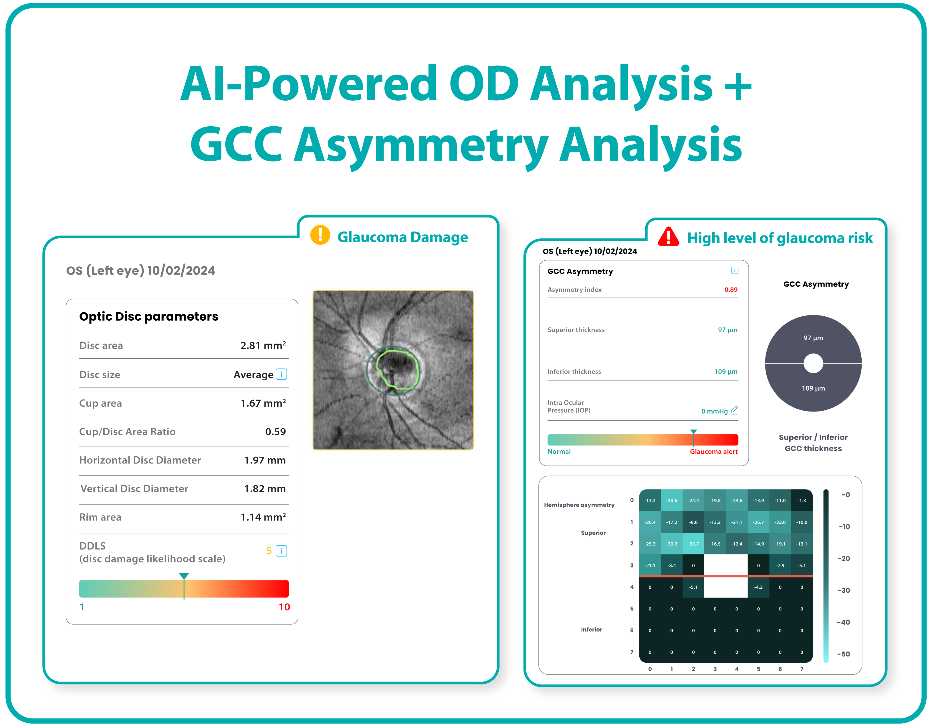
Altris AI GLAUCOMA MODULE


Altris AI is an artificial intelligence platform for OCT analysis, apparently capable of detecting more than 70 retinal pathologies and biomarkers, the highest among such technologies worldwide.

Altris AI has recently launched an advanced glaucoma "Optic Disc Analysis module" for OCT scans.

Traditionally, OCT results are dependent on normative databases pre-loaded in the machine. These databases can be insufficient or skewed for populations not usually seen in the region. For example, the machine can have more caucasian datasets, even though the population being evaluated is Asian or African. 

The new module evaluates optic disc parameters using OCT, providing personalized assessments by accounting for individual disc sizes and angle of rim absence. This tailored approach eliminates reliance on normative databases, making evaluations more accurate and patient-specific.

Key parameters evaluated by Altris AI’s Optic Disc Analysis include:

  1. Disc area
  2. Cup area
  3. Cup volume
  4. Minimal Cup depth
  5. Maximum Cup depth
  6. Cup/Disc area ratio
  7. Rim Absence angle
  8. Disc-Damage Likelihood Scale (DDLS)

OCT Test for Glaucoma - altris

Altris AI’s platform assigns a severity score for optic disc damage on a scale from 1 to 10, offering valuable insights into glaucomatous changes. Furthermore, it enables cross-evaluation across different OCT systems, allowing practitioners to analyze both macula and optic disc pathology, even when data originates from multiple OCT devices.

By combining GCC Asymmetry (a program developed by Altris AI previously, designed to detect early risk of glaucoma), and Advanced Optic Disc analysis for glaucoma, this technology enables eye-care practitioners to make faster evaluations and explore a wider range of treatment options. This streamlined approach empowers users with timely, actionable data, ultimately improving patient outcomes and care.

This innovation not only reduces false positive referrals but also enhances early detection and treatment planning—ensuring better outcomes for patients and optimizing healthcare resources.

REFERENCE:

https://aiineyecare.com/altris-ai-introduces-advanced-optic-disc-analysis-for-glaucoma/#

Wednesday, June 18, 2025

PreserFlo Microshunt occlusion

Bonatti et al have presented the successful resolution of early fibrin occlusion of PreserFlo MicroShunt with Nd:YAG laser treatment. It is regarded as the first reported case of such a nature.

The PreserFlo microshunt device is indicated in patients with moderate or advanced open-angle glaucoma who require further intraocular pressure (IOP) reduction to prevent disease progression. 

The 70-mm lumen implant redirects aqueous fluid from the anterior chamber to the subtenon space. The microshunt works immediately after its implantation, significantly reducing the IOP, typically in the single digits on the initial post-operative period.

Obstruction of the implant lumen can reduce the aqueous flow through the shunt and prevent this pressure reduction.

The authors performed a PreserFlo MicroShunt implant with phacoemulsification and acrylic intraocular lens implantation on the left eye of a 64-year-old male patient with primary open-angle glaucoma (POAG).

On the first postoperative day, he presented with an uncomfortable left eye with best corrected visual acuity (BCVA) of 6/15, mild corneal edema, and a centered intraocular lens. No filtering bleb was observed and IOP in that eye was 39 mmHg. The intracameral portion of the PreserFlo MicroShunt was in a good position in the anterior chamber, but fibrin strands were observed in the AC going toward the internal ostium of the shunt.


Apparently, the fibrin deposition was limiting drainage through the shunt. The authors used neodymium-doped yttrium aluminum garnet (Nd:YAG) laser to clear the obstructing material. 

The material was lysed under direct visualization using 10 single shots with a power of 1.1 mJ. 

Upon breaking the material, the authors immediately observed a fibrin strand being aspirated into the shunt lumen, indicating patency of the implant. However, 10 minutes after the procedure, the filtering bleb remained flat, and the IOP was 38 mmHg, without any evidence of fibrin in the anterior chamber.

It was suspected that the previously noted fibrin strand was trapped inside the shunt lumen and the patient underwent another laser procedure. 

With the aid of a Ritchie lens, the authors lasered the midsection of the shunt's lumen under the conjunctiva. Two single shots were given with a power of 1.0 mJ until gas bubbles were observed inside the lumen. After 15 minutes, the patient was symptomatically comfortable, an elevated filtering bleb was observed, and the IOP was 12 mmHg. 

At the 1-week postoperative visit, there was no evidence of fibrin in the AC, BCVA was 6/6 and IOP was 4 mmHg. 

At his last visit, 12 months after surgery, his IOP was 15 mmHg with brimonidine-timolol combination twice daily.

REFERENCE:

Bonatti R, Hodgson K, Nicolela M. Successful resolution of early fibrin occlusion of PreserFlo MicroShunt with Nd:YAG laser treatment. Can J Ophthalmol. 2025 Jun;60(3):e482-e485. doi: 10.1016/j.jcjo.2025.01.003. Epub 2025 Feb 6. PMID: 39870357.

Sunday, June 8, 2025

PROSTAGLANDINS & RISK OF UVEITIS


Prostaglandin Analogues (PGAs) have often been regarded as pro-inflammatory and contraindicated in situations where there is risk of inflammation, such as neovascular glaucoma (NVG).

Chauhan et al have published a study to evaluate the risk of uveitis among patients starting on topical glaucoma therapy.

The retrospective study utilized the Sight Outcomes Research Collaborative (SOURCE) Ophthalmology Data Repository data from 10 health systems contributing to the SOURCE data repository. 

Adult glaucoma patients who were recently started on topical glaucoma therapy were included in the study. The anti-glaucoma medications included prostaglandin analogs [PGAs], beta-blockers [BBs], alpha agonists [AAs], and carbonic anhydrase inhibitors [CAIs].

Patients with pre-existing documentation of uveitis were excluded.

The main outcome measure was the incidence of uveitis within 3 months of initiating therapy with different topical glaucoma medications.

The study included 67517 patients who were newly prescribed a topical glaucoma medication. 

A total of 567 patients (0.87%) developed uveitis within 3 months of initiating the therapy. 

The incidence of uveitis was 1.95%, 1.68%, 1.63%, and 0.32% for users of BBs, CAIs, AAs, and PGAs, respectively.

After adjusting for sociodemographic factors, individuals using topical BBs, AAs, and CAIs had significantly higher odds of developing uveitis versus those using PGAs (P < 0.001 for all comparisons).

The study concluded that the use of PGAs was not associated with higher odds of developing uveitis compared with other classes of topical glaucoma medications.

REFERENCE:

Chauhan MZ, Elhusseiny AM, Marwah S, Sallam AB, Stein JD, Kishor KS; SOURCE Consortium. Incidence of Uveitis Following Initiation of Prostaglandin Analogs versus Other Glaucoma Medications: A Study from the Sight Outcomes Research Collaborative Repository. Ophthalmol Glaucoma.2025;8:126-132.


RETINITIS PIGMENTOSA AND GLAUCOMA

Retinitis pigmentosa (RP) is a rare inherited retinal disorder, characterized by night-blindness and ultimately loss of vision due to involv...