

Ophthalmologists often debate the advantage of structural versus functional studies to diagnose glaucoma. There have also been efforts to combine the two and come up with a structure-function index. The crux of these observations is the need to develop mechanisms which would identify glaucomatous changes at the earliest. Since, a sub-group of patients may manifest structural changes earlier than functional changes and vice-versa, (“the structure-function disassociation”) it has become controversial as to which method is preferable.

The need to develop a singular diagnostic method utilizing structural and functional tests is based on the above mentioned probability of structure-function disassociation. Since, one type of test may be normal in an individual, it is required to perform another type of test to confirm the diagnosis. According to Elliot Kirsten, “Our efforts to coalesce ocular physical measurements with functional values seem to point to our desire to define glaucoma in one simple value. While the effort itself has already shed great benefits to our understanding of this complex disease, it ultimately seems unlikely that we will ever be able to adequately define glaucoma in terms of a singular value”.
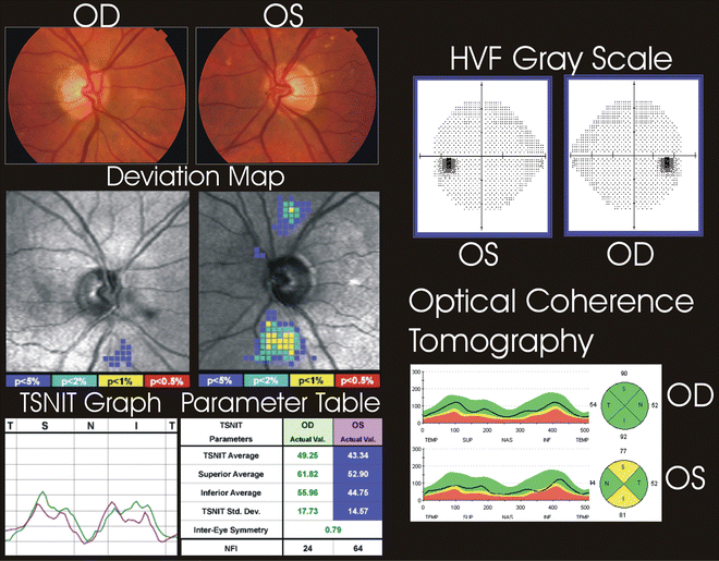
A structural measure for glaucoma assessment refers to measurements of the neuroretinal rim, retinal nerve fibre layer (RNFL) and the ganglion cell layer in the macula. The initial methods to study glaucomatous changes at the disc and parapapillary retina started with the development of the direct ophthalmoscope by Hermholtz in 1851. Using this instrument, ophthalmologists were able to study the optic disc and detect changes in its structure. Usually, ophthalmologists resorted to diagrams to denote the cup, disc and other changes. This is still used in a large number of practices. However, these findings are prone to intra- and inter-observer differences. Fundus photography, especially stereo-photographs have a better chance of being consistent among observers. Newer digital platforms are available to capture the images. Study of the disc with instruments such as GDx and Heidelberg Retinal Tomograph (HRT) have brought another dimension to these methods. Unfortunately, these instruments are expensive and their cost-benefit efficiency is probably limited when regarded as a whole in glaucoma diagnostic techniques.
The development of optical coherence tomography (OCT) provided us a vast data set, allowing us to seek out specific retinal measurements that are most highly specific for the diagnosis and observation of glaucomatous progression. OCT provides information regarding the disc, surrounding RNFL and other layers of the retina. Advanced segmentation techniques are giving us important and precise information of the layers most affected in glaucoma. OCT-angiography is also being used to demonstrate areas of retinal loss. Another technique being developed is the Detection of Apoptosing Retinal Cells (DARC). This technique pin-points the apoptosing retinal ganglion cells and gives a true picture of cellular damage in glaucoma patients.
Functional tests are largely based on visual field analysis. Other tests such as Visual Evoked Potential (VEP), pattern electroretinography (pERG), and Relative Afferent Pupillary Defect (RAPDx) have not been much used in common clinical practices and are usually limited to research purposes. The first record of a visual field defect is found in Hippocrates’ description of a hemianopia from the late fifth century B.C. Later, Ptolemy (150 B.C.) quantified the visual field and noted its circular form. Perimetry has now advanced in terms of better hardware and software.
Standard automated perimetry is still the gold standard for detecting functional change. A post-hoc analysis of the Early Manifest Glaucoma Trial (EMGT) by Öhnell et al. has shown that after a median follow-up of 8 years, eyes with a visual field defect at baseline were more than 4 times more likely to have progression first detected as further worsening of the visual field (VF) compared with progression on optic disc photographs. However, since the EMGT was an old study, the findings may not be accurate in the current scenario. Newer imaging systems are providing better documentation of RNFL thinning compared to photographs.
Thus, the question being raised is whether structure or functional tests are more reliable. It is to be highlighted that we need both set of tests to diagnose the complex disease called glaucoma. It is not the deficiency of either type of test in detecting glaucomatous changes, but it is the varied pathogenesis of glaucoma in different individuals which produces this structure-function disassociation. Hence, we may conclude by this sobering thought that no single technique is presently available to diagnose glaucoma with significant sensitivity and specificity in all individuals.

