Tuesday, May 19, 2020

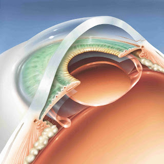
The eyeWatch glaucoma device


Glaucoma is a multifactorial disease characterized by progressive damage to the retinal ganglion cells (RGCs) and its axons. The management of this condition is presently restricted to control of intraocular pressure (IOP) to such levels that progression can be reduced or even halted. Management consists of pharmacological methods, laser procedures, and surgery. Conventionally, surgery was restricted to some type of filtering procedure which allowed aqueous to drain from the eye. However, trabeculectomy failure rates can be a significant problem in certain series of patients. Over the last few years, glaucoma drainage devices (GDDs) have been introduced to overcome the myriad complications seen with trabeculectomy. However, these methods too are far from successful in all patients.

One of the major problems with glaucoma filtering surgeries and also GDDs is that of hypotony which occurs in the immediate post-operative period. This occurs either due to over-filtration or possibly the ciliary shock mechanism, which causes less aqueous humor to be produced in the early post-operative phase. While this problem is not so significant in trabeculectomy, as surgeons have found methods to control that by releasable sutures, padding the eye and so on, this complication is more significant following the use of GDDs. Some GDDs have a rip-cord in their drainage tube which can be removed after a few days of surgery. Others employ sutural ligation of the tube to prevent early postoperative hypotony. The drawbacks of such flow-restricting techniques are the lack of precision and predictability in efficiently controlling IOP in the early postoperative phase, which may result in volatile IOP with initial hypertensive and subsequent hypotensive phase.

Recently, a new method to control aqueous drainage following the implantation of GDDs has been developed in Switzerland. This device is known as the eyeWatch. Developed by Rheon Medical, Lausanne, Switzerland, the device can adjust the aqueous outflow and thus, better control IOP. The device consists of a deformable silicone tube which connects the anterior chamber to any GDD. Rheon Medical also has an in-house plate valve known as the eyePlate.


The tube is compressed by rotating a magnetic disc on a shaft eccentric to its axis of symmetry. This compression makes changes in the cross-sectional area of the tube and thus, changes the fluidic resistance. This in turn, alters the aqueous outflow through the anterior chamber. The magnetic disc is adjusted by a non-invasive external control unit known as the eyeWatch Pen. The Pen is kept 1-2 mm from the device and dialed from 0 to 6, indicating fully open to fully closed respectively.

The steps in the surgical procedure to implant eyeWatch are available at the following website:

In short, a GDD is first sutured to the sclera. A bed in the sclera is created using the eyeWatch marker. Subsequently, a 25-G needle is used to enter the anterior chamber. An anterior chamber maintainer is useful. The nozzle of the eyeWatch is stiff and short. This avoids damage to the corneal endothelium. The eyeWatch is sutured to the sclera. Subsequently, the tube of the GDD is cut to an appropriate length and attached to the eyeWatch. Finally, a 7mmx7mm pericardial patch graft is sutured to the sclera to cover the device and prevent tube erosion. No anti-metabolites are required in this surgery.


In a study of 15 patients implanted with the eyeWatch, mean baseline pre-operative IOP of 26.2±6.8mmHg reduced to 11.9±2.8mmHg at 12 months (P<0.001). The mean number of glaucoma medications decreased from 3.0 ±0.7 before surgery to 0.8 ±0.9 at last visit (P<0.001). The success rate was 40% for complete success (IOP between 6-18 mmHg, with a 20% reduction from baseline, without medications) and 93% for overall success at last follow-up. Complication rate was 7%. The complications affecting 4 patients included 2 cases of conjunctival wound leak (Seidel sign positive) and 2 cases of choroidal detachment.


A few questions are raised regarding the procedure. One, is the magnet affected during MRI? The study mentioned above did not find any interference of the imaging technique by eyeWatch. Although, all patients (n=4) who underwent MRI required readjustment of the eyeWatch settings following the MRI. The second question is that the device does not address the main causes of GDD failure, that is, fibrosis around the implant. Another question which can be asked is whether the presence of a magnet inside the human eye would affect the physiology of not just the eye, but also the body. There are conflicting reports regarding the ability of the human body to sense magnetic fields. Birds and bats are classic examples of using the earth’s magnetic field to navigate. Although humans probably do not need those cues for their sense of direction, the long term effects of such devices in the eye are unknown. It should also be cautioned that GDDs are often used in patients with advanced glaucoma, a group in whom vision and visual fields are already compromised.       

Long-term results of this device shall be awaited by glaucoma surgeons, as this could prove to be an important development in IOP adjustment following successful glaucoma surgery. 


Sunday, May 10, 2020

CLEAR-LENS EXTRACTION FOR PRIMARY ANGLE CLOSURE DISEASE


Dr Tooba, Dr Iram, Dr Ghuncha, Dr Anwar, Dr Naeem

The lens plays an important role in the development of primary angle closure disease (PACD). This concept was first given by Priestley Smith in 1891.

Conventionally, PACD is managed by pharmacological means or laser procedures. Usually, these two modalities are sufficient to control intra-ocular pressure (IOP). However, ocassionally these techniques are not sufficient to bring IOP down to target pressure levels. In such situations other methods have to be attempted.

Over the years, cataract extraction has become a second-line procedure to bring down IOP in such cases. A number of reports have been published regarding successful outcomes following cataract surgery (ECCE or phacoemulsification) in eyes with PACD. 

However, the new modality of Clear-Lens Extraction (CLE) for managing PACD has become a subject of debate. Unlike cataract surgery this technique involves removal of a clear lens where visual symptoms are minimal. A number of studies such as the landmark EAGLE study and those by Tham, Dada and Man have shown that CLE could be a useful procedure to deal with PACD.

The studies mentioned above have thrown up a lot of positive as well as negative aspects regarding the conduct of the studies themselves and the inferences coming out of them.

In such a scenario we performed a review of CLE in cases of PACD. The article will be published in a forthcoming issue of US Ophthalmic Review. The ahead of publication epub is available at the following link:

https://www.touchophthalmology.com/clear-lens-extraction-in-primary-angle-closure-disease-pros-and-cons/ 


Friday, May 1, 2020

HAR GOBIND KHORANA: THE HUMBLE GENIUS


[COVID-19 SERIES#4]


In these times of the COVID-19 pandemic, genetic manipulation and testing are increasingly in focus. On one hand there are rumors that the causative Corona virus is man-made, while on the other there are efforts to crack the genetic code of the virus and develop a vaccine as soon as possible. In the backdrop of this situation one name which comes to mind is that of Dr. Har Gobind Khorana, the first researcher of Indian origin to win the Nobel Prize for Physiology or Medicine.


A Google doodle honoring the researcher


Dr. Khorana was born on 9th January, 1922 in the village of Raipur in Multan (present Pakistan), where his father was working as a clerk. His family was the only literate among the 100 or so living in the village. After schooling, he was offered a scholarship at Punjab University, Lahore to study chemistry. However, he was so shy that he refused to attend the scholarship interview and almost took up English. Nevertheless, he got the chemistry scholarship and completed his Bachelor course in 1943 and Masters in 1945.


Soon thereafter, he won a scholarship to study organic chemistry at the University of Liverpool. He obtained his Ph.D. in 1948 under the supervision of Roger J.S. Beer. Subsequently, he went to Zurich, Switzerland for post-doctoral research. There, he joined the Eidgenössische Technische Hochschule (ETH) working under Vladimir Prelog, a chemist who won a Nobel Prize in 1975 for his work on stereochemistry. However, within a year Gobind ran out funds and secretly lived in the lab to avoid expenses. Subsequently, he was forced to return to India.


However, in India the young scientist became jobless. But then Lady Luck smiled again and he won a three-year fellowship with Alexander Todd at Cambridge University. While at Cambridge, Gobind was exposed to Sanger's exciting advances in protein sequencing, Perutz's and Kendrew's breakthroughs in protein crystallography, and Todd's work on the chemical structures of nucleic acids. This innovative environment drew Dr. Khorana, a synthetic organic chemist, to the newborn field of Molecular Biology. The iconic Cambridge University would in 1953 be the site where James D. Watson and Francis H. C. Crick would discover the double-helix structure of DNA.


In 1952, Dr. Khorana had the exciting prospect of setting up his lab by the British Columbia Research Council in Vancouver. There, he started work on nucleotides and nucleic acids. He used a carbodiimide (dicyclohexylcarbodiimide) to form pyrophosphate bonds, which eventually led to the first synthesis of coenzyme A and ATP. 


In 1952 he married Esther Elizabeth Sibler, whom he had first met in Switzerland. The couple had three children, Julia Elizabeth, Emily Anne, and Dave Roy. He credited her with bringing a “consistent sense of purpose into my life at a time when, after six years’ absence from the country of my birth, I felt out of place everywhere and at home nowhere.”


In 1960 Khorana accepted a position as co-director at the Institute for Enzyme Research at the University of Wisconsin at Madison, USA. He became a professor of biochemistry in 1962 and was named Conrad A. Elvehjem Professor of Life Sciences at UW–Madison. Most of his significant work which led to the Nobel Prize were performed here. In 1966 he accepted the US citizenship.


He generated synthetic oligonucleotides and amplified these molecules biosynthetically with DNA polymerase. Using the oligonucleotide CUCUCU, he discovered that the triplets CUC and UCU encode the amino acids leucine and serine, respectively. This work corroborated the studies of Marshall Nirenberg, who had previously shown that a UUU triplet encoded a phenylalanine residue. Gobind, with his characteristic humility, would always note that Nirenberg's work inspired his own investigations of the genetic code.


In 1968 he won the Nobel Prize for Physiology or Medicine with Marshall W. Nirenberg and Robert W. Holley for research that showed the order of nucleotides in nucleic acids, which carry the genetic code of the cell and control the cell's synthesis of proteins. He would often rent a room or cottage without a phone, radio, or TV so that he could think and write without distraction. His wife, Esther was forced to drive an hour to inform Dr. Khorana that he had won the Nobel Prize.


In 1970 Dr. Khorana joined the Massachusetts Institute of Technology as the Alfred P. Sloan Professor of Biology and Chemistry. After a long and fruitful career at MIT he would retire in 2007.


While at MIT, using DNA ligases, Gobind's lab assembled the coding region of the gene for the alanine tRNA. By 1976, Gobind had added the required regulatory elements needed to express the gene in a living bacterial cell and demonstrated that the synthetic tRNA functioned identically to the naturally expressed gene. This seminal work defined the conceptual and technical framework for biotechnology and, nearly 45 years later, is still the strategy used to assemble synthetic genes and genomes.


Gobind's mentorship involved rigorous intellectual training and hard work. It did not matter if none of the experiments worked (at least for the impossible problems!), but complete “24/7” engagement was expected. One possibly apocryphal story involved Saturday morning donuts that Gobind would bring to the lab. Rumor had it that he had identified everyone's favorite type of donut and would bring only one each. At the end of the day, he would check the remaining donuts and determine who had come in over the weekend!


Dr Khorana won eminent honors from all over the world. Among others, he was awarded the Louisa Gross Horwitz Prize, Lasker award, Willard Gibbs Medal, Gairdner Foundation Annual Award, Paul Kayser International Award of Merit and the National Medal of Science. An unassuming man to the last, he was sent numerous letters for his National Medal of Science award, which were unanswered. Ultimately, a White House representative tracked him to a conference where he got the assurance from Dr. Khorana that the researcher would attend the felicitation ceremony. 


Dr. Har Gobind Khorana passed away on 9th November 2011, in Concord, Massachusetts, at the age of 89. In 2002, the government of India was requested to grant him its highest civilian award, the Bharat Ratna. Unfortunately, the country is yet to pay this colossal legend a worthy obituary.


CARONIA GLAUCOMA CARD

  The Amsler Grid chart, a commonly used method for detection of macular pathologies, has been utilized in detection of visual field (VF) de...