RHO KINASE INHIBITORS IN GLAUCOMA
Guest author
RAMSHA ANWAR
Ajmal Khan Tibbiya College, Aligarh, India

INTRODUCTION:
Rho Kinase is a serine/threonine protein
kinase involved in the regulation and modulation of cell shape and size via its
action on the cytoskeleton.
FUNCTIONS
OF RHO KINASE:
As
downstream effectors of Rho GTPase, Rho Kinases are involved in calcium
independent regulation of smooth muscle contraction.
(A small
GTP-binding protein, called Rho, acts on ROCKs downstream, to produce certain
changes in the tissues)
Rho
Kinases are also linked with:
(1) the control of cytoskeleton dynamics
(2) actinomyosin contractile forces
(3) cell adhesion
(4) cell stiffening
(5) extracellular matrix and re-organization
(6) cell morphology.
(1) the control of cytoskeleton dynamics
(2) actinomyosin contractile forces
(3) cell adhesion
(4) cell stiffening
(5) extracellular matrix and re-organization
(6) cell morphology.

The
above mentioned factors regulate aqueous humor outflow via the trabecular
pathway resulting in lowered intra-ocular pressure (IOP).
Rho
Kinases (ROCK) exist in 2 isoforms: ROCK 1 and ROCK 2.
ROCK
1 is located on chromosome 18 and contains 1354 aminoacids.
ROCK 2 is located on chromosome 12 and encodes a 1388 aminoacid product.
ROCK 2 is located on chromosome 12 and encodes a 1388 aminoacid product.
In
humans ROCKs are expressed in majority of tissues, including trabecular
meshwork and ciliary muscle cells.
Structurally,
ROCKs are composed of 3 major domains:
- An N-terminal kinase domain
(which phosphorylates protein targets).
- A C-terminal kinase domain (which
limits kinase activity via intramolecular interactions)
- A coiled-coil Rho-binding domain
(which facilitates the switch from the inactive to active conformation).
On
binding to Rho, the catalytic activity of ROCKs is moderately enhanced.
ROCKs
inhibit myosin light chain phosphatase thereby mediating actin cytoskeletal
changes. These directly affect the contractile properties of trabecular
meshwork outflow tissue.
ROCK
promotes the assembly of actin stress fibers and focal adhesions.
ROCK
also regulates cell contraction and motility.
Inhibition
of ROCK improves aqueous outflow through the trabecular meshwork resulting in
lowered IOP.

RHO
KINASE SIGNALING PATHWAY:
- Rho Kinase is a downstream
effector of the RhoA protein, a small GTPase.
- GTPase interchange between two
conformations (a) A Guanosine triphosphate (GTP)-bound ACTIVE conformation and
a Guanosine Diphosphate (GDP)-bound INACTIVE conformation.
- A number of factors control
GTPase activation. These regulators include:
·
Guanine
nucleotide Exchange Factors (GEFs)
·
GTPase
Activating Proteins (GAP)
·
Guanine
nucleotide Dissociation Inhibitors (GDIs)
·
Myosin
Light Chain
·
LIM
kinase
The above mentioned substrates interact to control actinomyosin contractility, membrane permeability, cellular adhesion, cell stiffening, cell morphological changes, extracellular matrix organization and DNA synthesis.
EFFECTS OF RHO KINASE INHIBITORS
AS IOP LOWERING AGENTS


No new class of anti-glaucoma drugs
has been introduced since 1996, when FDA approved latanoprost.
Therefore, this group of Rho
Kinase inhibitors are an exciting new class of drugs available to lower IOP.
Two drugs are commonly available
commercially. These include: Ripasudil (K-115) and Netarsudil (AR-13503).
DRUGS
|
RIPASUDIL
|
NETARSUDIL
|
COMMERCIAL
NAME
|
Glanatec (Derivative of Fasudil)
|
Rhopressa
|
CHEMICAL
FORMULA
|
C15H18FN3O2S
|
C28H27N3O3
|
IUPAC
NAME
|
4-fluoro-5-(((2S)-2-methyl-1,4-diazepan-1-yl)sulfonyl)isoquinoline
|
(4-((1S)-1-(Aminomethyl)-2-(isoquinolin-6-ylamino)-2-oxoethyl)pheny)methyl2,4-dimethylbenzoate
|
DURATION
IT TAKES TO LOWER IOP AFTER INSTILLATION
|
2
hours
|
2
hours
|
DOSAGE
|
0.4%
drug instilled twice daily
|
0.02%
solution used once daily
|
SIDE
EFFECTS
|
Conjunctival
hyperemia
Conjunctival
haemorrhage
(Hyperemia
usually resolves spontaneously over few hours)
ROCK
inhibitors usually lower BP and vascular resistance.
ROCK
inhibition reduced the intraocular penetration of concurrent timolol
instillation (presumably by increasing the elimination through the dilated
conjunctival vasculature).
|
Conjunctival
hyperemia
Cornea
verticillatae (Usually asymptomatic and did not reduce visual function)
Pain
at instillation site
Conjunctival
haemorrhage
|
MECHANISM:
ROCK inhibition lowers IOP by:
- Increasing aqueous humor outflow.
- Reducing aqueous humor production.
- Decreasing episcleral venous pressure (EVP). This can be understood by the following equation=
IOP= EVP + 1/ C(F-U)
Here, C= Facility of outflow.
F= Formation of aqueous humor.
U= Resorption rate of aqueous
humor.
IOP= Intra-ocular pressure.
As the metabolic pathway of Rho
Kinase controls many aspects of cell morphology, when it is inhibited following
changes occur:
- Change of cell shape in the trabecular meshwork.
- Change in actin cytoskeletal
structure
- Rounding of cell bodies
- Disruption of actin production.
All
these changes allow greater outflow of aqueous humor through the trabecular
meshwork which ultimately results in decreased IOP.
NOR-EPINEPHRINE
TRANSPORT INHIBITION:
Many
ROCK inhibiting drugs chemically include a nor-epinephrine transport (NET)
inhibition component.
This
nor-epinephrine inhibitor helps to reduce aqueous production and decreases EVP.
Nor-epinephrine
transport inhibition lowers aqueous humor production by (i) vasoconstriction
(ii) reducing blood flow to the ciliary processes.
Aqueous
humor production may be reduced by 20-23% by nor-epinephrine transport
inhibition.


NEUROPROTECTION:
ROCK
inhibitors increase blood flow in the optic nerve head by relaxation of the
vascular endothelial smooth muscle. Nitric oxide synthase inhibitor induced
impairement of optic nerve blood flow was also prevented by ROCK inhibition.
ROCK
inhibitors influence neuron survival and axon regeneration. Fasudil protected
against glutamate-related excitotoxicity in the retina and better preserved
cells of the ganglion cell layer on exposure to N-methyl-D-aspartate.
CLINICAL
TRIALS:
Phase
1,2,3 clinical trials of netarsudil were conducted in more than 2000 patients.
0.02% once daily in the evening was found to be the most efficacious and well tolerated
dosing regimen. The phase 2 clinical trial included comparison with latanoprost
and four phase 3 trials comparing netarsudil with timolol.
When
netarsudil and latanoprost were compared, IOP reductions were similar [-5.8
mmHg in Netarsudil group vs -5.9 mmHG in the latanoprost group]. Among all
patients netarsudil was 1 mmHg less effective than latanoprost and did not meet
the statistical analysis for non-inferiority of netarsudil to latanoprost
criteria.
Rocket
1,2,4 phase 3 trials compared netarsudil to timolol. All studies showed
comparable IOP reduction with both drugs.
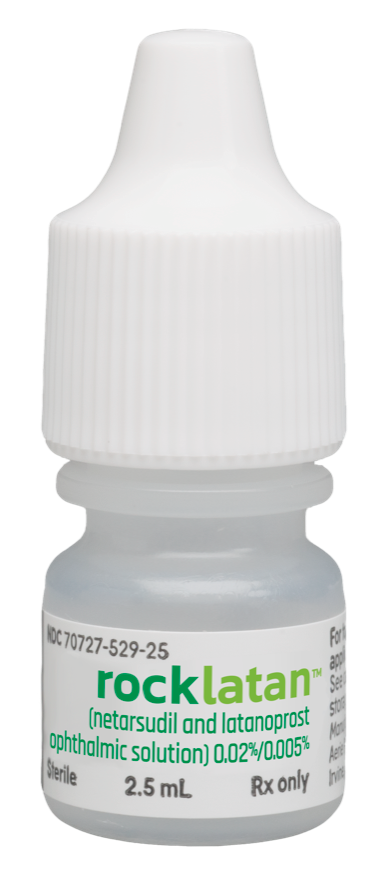
A
fixed-dose combination of netarsudil and latanoprost (Roclatan) has been tested
with individual components in two clinical studies (Mercury 1 and Mercury 2).
Mercury 3 is an ongoing trial comparing Roclatan and Ganfort.
Mercury
1 & 2 have shown 1-3 mmHg greater IOP lowering with Roclatan compared to
the individual components. IOP reductions of atleast 30% were achieved in 65%
of patients treated with Roclatan, compared to 40% when individual components
were used.
Roclatan
treated patients achieved 16 mmHg or less IOP in 61% cases, while ≤ 14 mmHg was
achieved in 33% cases. This was comparable to those treated with individual
components who achieved similar results in 40% or less and 15% or less patients
respectively.


CONCLUSION:
Use
of Rho Kinase inhibitors in treating glaucoma has been proven to be of benefit.
Although there is evidence showing that Rho Kinase inhibitors are related to
increased conjunctival hyperemia.


No comments:
Post a Comment