IRIDOCORNEAL DYSGENESIS

Individuals with malformations of structures of the anterior segment of
the eye frequently develop elevated intraocular pressure and glaucoma. This post takes a brief figurative look at some of these conditions which lead to developmental glaucomas.
1. POSTERIOR EMBRYOTOXON: Characterized by anterior displacement of Schwalbe's Line.


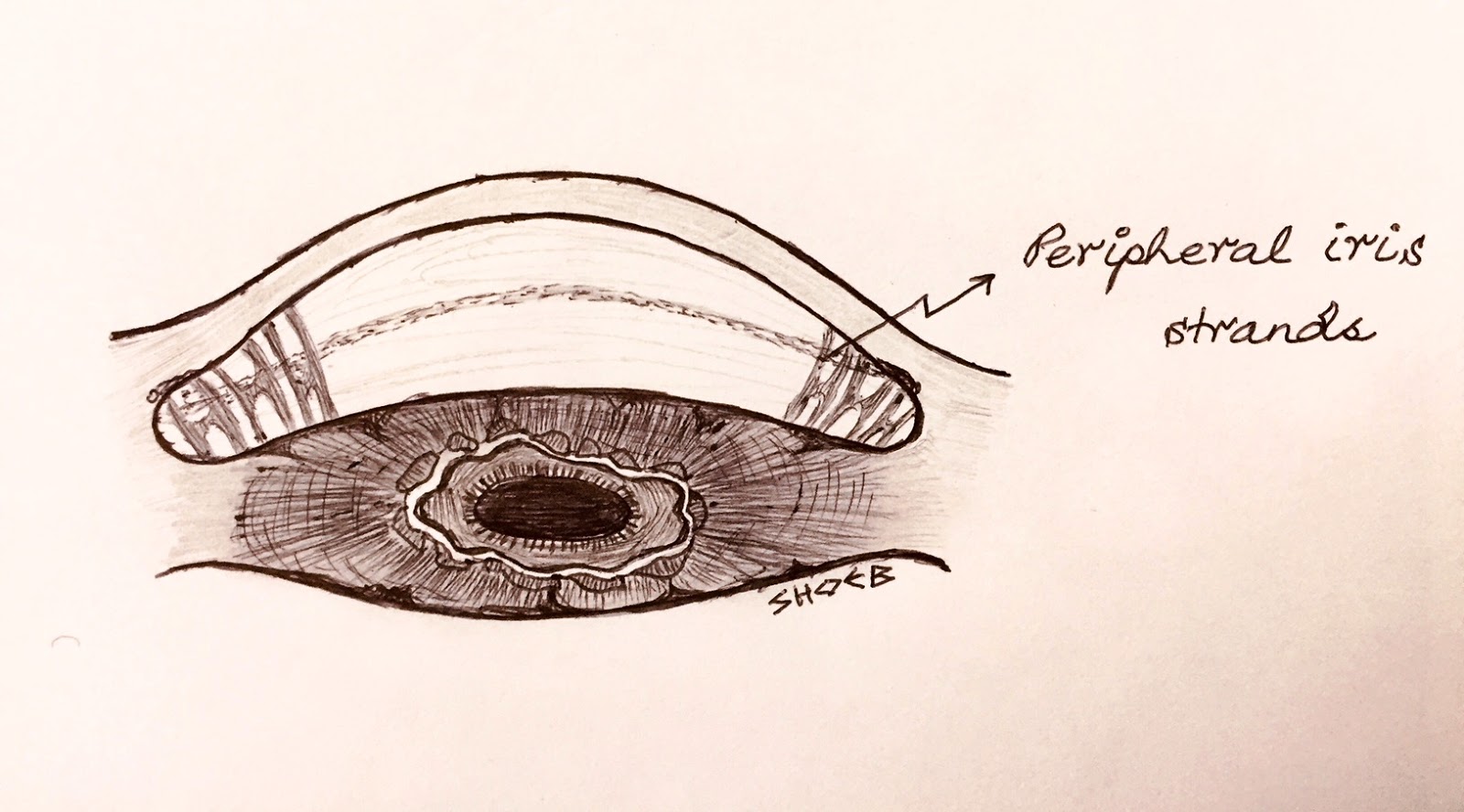
2. AXENFELD ANOMALY: Characterized by posterior embryotoxon and peripheral iris strands.
3. RIEGER ANOMALY: Characterized by iris hypoplasia, correctopia and polycoria.


When Rieger anomaly is associated with systemic features such as: dental and facial defects e.g. maxillary hypoplasia; umblical abnormalities and pituitary involvement, it is called RIEGER SYNDROME.
Association of Axenfeld anomaly and Rieger anomaly together is known as: AXENFELD-RIEGER SYNDROME.

4. PETER SYNDROME: Characterized by central or paracentral corneal opacity, absent Descemet's membrane and endothelial layers. There are also iridocorneal adhesions from the border of the central corneal opacity. Peter's anomaly is of 2 types. Type 1 is unilateral, while Type 2 is bilateral in about 60% cases with lens adherent to the posterior surface of the cornea due to lack of separation between the 2 layers. Type 2 is also associated with systemic features such as: short stature, mental retardation, abnormal ears, cleft lip and/or palate, defects of extremities, genitourinary system defects, cardiovascular anomalies, and gastrointestinal defects.

Some good articles on the subject can be accessed from here:


Thanks to Prof Hommer for the correction.
ReplyDeleteWow very impressive, keep up the great work
ReplyDeleteThank you very much
Delete